Degenerative Changes

There is sclerosis, scoliosis, and discordance of bone mineral density seen in this Hologic lumbar spine scan. Such degenerative changes will overestimate the bone mineral density in the lumbar spine and make it less useful in followup. An additional site such as the 1/3 wrist should be added in a patient with degenerative changes in the lumbar spine.
There are severe degenerative changes in the right femoral head which extend into the femoral neck. This results in discordance in the bone mineral density between the femoral neck and total hip. In this case, the left hip had been replaced, so the right hip was scanned.

There are degenerative changes in the distal wrist. The 1/3 radius is unaffected by degeneratie changes. The presence of degenerative changes can obscure usual landmarks.
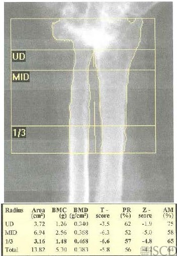
Degenerative changes are common internal artifacts. These panels show degenerative changes in the spine, hip, and wrist. The presence of degenerative changes, depending on location, may make the scan less useful in diagnosis (accuracy) and follow-up (precision)
Sarah L Morgan, MD, RD, CCD, The University of Alabama at Birmingham
• Banks, L.M., et al., Effect of degenerative spinal and aortic calcification on bone density measurements in post-menopausal women: links between osteoporosis and cardiovascular disease? Eur J Clin Invest, 1994. 24(12): p. 813-7.
• Burger, H., et al., Association of radiographically evident osteoarthritis with higher bone mineral density and increased bone loss with age. The Rotterdam Study. Arthritis Rheum, 1996. 39(1): p. 81-6.
• Ichchou, L., et al., Relationship between spine osteoarthritis, bone mineral density and bone turn over markers in post menopausal women. BMC Womens Health, 2010. 10: p. 25.
• Knight, S.M., E.F. Ring, and A.K. Bhalla, Bone mineral density and osteoarthritis. Ann Rheum Dis, 1992. 51(9): p. 1025-6.
• Liu, G., et al., Effect of osteoarthritis in the lumbar spine and hip on bone mineral density and diagnosis of osteoporosis in elderly men and women. Osteoporos Int, 1997. 7(6): p. 564-9.
• Orwoll, E.S., S.K. Oviatt, and T. Mann, The impact of osteophytic and vascular calcifications on vertebral mineral density measurements in men. J Clin Endocrinol Metab, 1990. 70(4): p. 1202-7.
• Peacock, D.J., et al., Lateral bone density measurements in osteoarthritis of the lumbar spine. Ann Rheum Dis, 1996. 55(3): p. 196-8.
• Preidler, K.W., et al., Dual-energy X-ray absorptiometric densitometry in osteoarthritis of the hip. Influence of secondary bone remodeling of the femoral neck. Acta Radiol, 1997. 38(4 Pt 1): p. 539-42.
• Pye, S.R., et al., Radiographic features of lumbar disc degeneration and bone mineral density in men and women. Ann Rheum Dis, 2006. 65(2): p. 234-8.
• Rand, T., et al., Impact of spinal degenerative changes on the evaluation of bone mineral density with dual energy X-ray absorptiometry (DXA). Calcif Tissue Int, 1997. 60(5): p. 430-3.
• Tenne, M., et al., Degenerative changes at the lumbar spine–implications for bone mineral density measurement in elderly women. Osteoporos Int, 2013. 24(4): p. 1419-28.
• von der Recke, P., et al., The impact of degenerative conditions in the spine on bone mineral density and fracture risk prediction. Osteoporos Int, 1996. 6(1): p. 43-9.
• Yu, W., et al., Influence of degenerative joint disease on spinal bone mineral measurements in postmenopausal women. Calcif Tissue Int, 1995. 57(3): p. 169-74.
• Reid, I.R., et al., The influence of osteophytes and aortic calcification on spinal mineral density in postmenopausal women. J Clin Endocrinol Metab, 1991. 72(6): p. 1372-4.