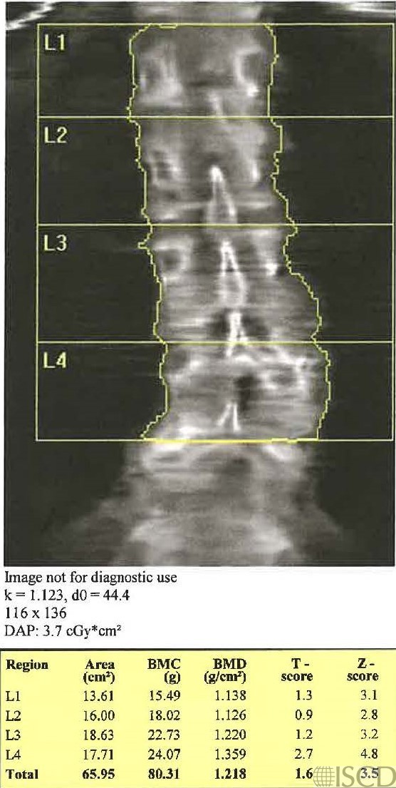
Legs Down vs Legs Up Spine Scan

The same patient was scanned within 5 minutes with the legs down and then the legs up on the block. This scan has the legs down.
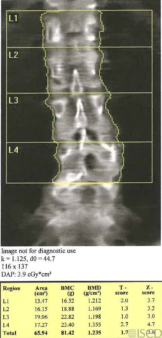
This scan has the legs up on the block. There is not a statistically significant difference in L1-L4 BMD using the 95% confidence intervals for the scanner.
Various authors have evaluated the effect of having the legs down, vs. on a block, for lumbar spine DXA scanning. Generally, the legs are placed on an appropriately sized block to flatten out the lumbar spine for scanning. Ikegami et al. evaluated spinal density with the patient supine or with the hips flexed to 90 degrees on a GE/Lunar Prodigy machine. The concluded that supine positioning slightly overestimated the bone mineral density compared to the flexed position, however, the positioning did not affect the detection of low bone mass (below). Lekamwasam and Lenora measured spine bone density on a Norland Eclipse XR in the supine vs flexed position. The concluded that the supine position did not alter the classification based on T-score vs. the flexed position. However, they concluded that hip flexion could be an important confounding factor when evaluating hip bone mineral density over time (below). Hwua measured spine bone density in the spine vs flexed position on a GE Lunar DPX and found a statistically increased spine bone density compared to flat positioning (below).
Sarah L Morgan, MD, RD, CCD, The University of Alabama at Birmingham
• Ikegami, S., et al., Clinical Implications of Hip Flexion in the Measurement of Spinal Bone Mineral Density. J Clin Densitom, 2016.
• Lekamwasam, S. and J. Lenora, Effect of hip flexion on the measurement of spinal bone mineral density in the Norland Eclipse XR. J Clin Densitom, 2005. 8(2): p. 183-6.
• Hwua, Y.-S., Difference in the spinal bone mineral denity measured with and without hips flexion. . J Clin Densitom, 2008. 11: p. 453.