Ostomy

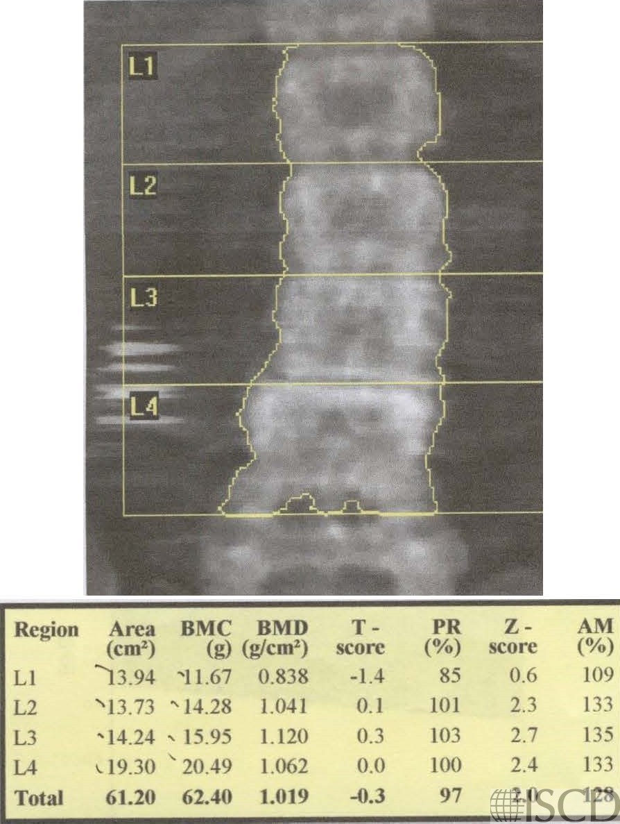
The DXA image shows an ileostomy that is lateral to L3-L4. The undo view showed that the internal artifact was removed from the soft tissue baseline.
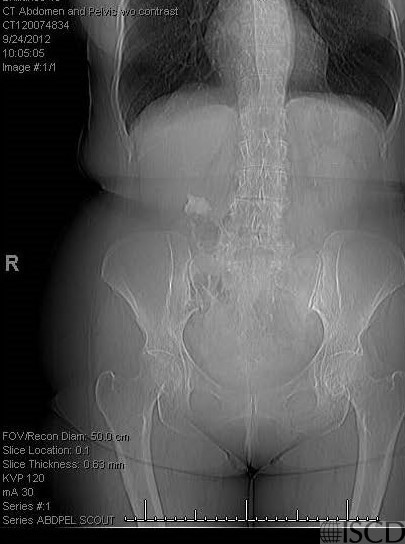
The accompany scout film from an abdominal CT scan also shows the ileostomy.
Sarah L Morgan, MD, RD, CCD The University of Alabama at Birmingham,
• Martineau, P., S. Bazarjani, and L.S. Zuckier, Artifacts and Incidental Findings Encountered on Dual-Energy X-Ray Absorptiometry: Atlas and Analysis. Semin Nucl Med, 2015. 45(5): p. 458-69.
• Bazzocchi, A., et al., Incidental findings with dual-energy X-ray absorptiometry: spectrum of possible diagnoses. Calcif Tissue Int, 2012. 91(2): p. 149-56.
• Bojinca, V., D. Opris, and M. Bonjinca, Artifacts and pitfalls in DXA scan images and interpretation. J Clin Densitom, 2012. 15(4): p. 486-487.