Ehlers-Danlos Syndrome

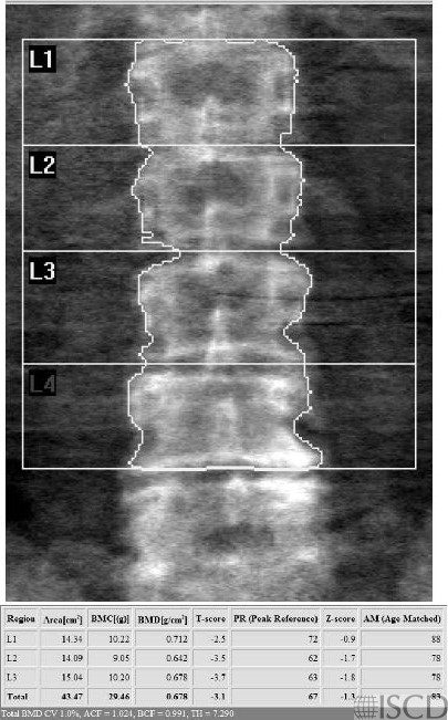
This is the Hologic lumbar spine DXA from a patient with hypermobile Elhers-Danllos syndrome showing osteoporosis.
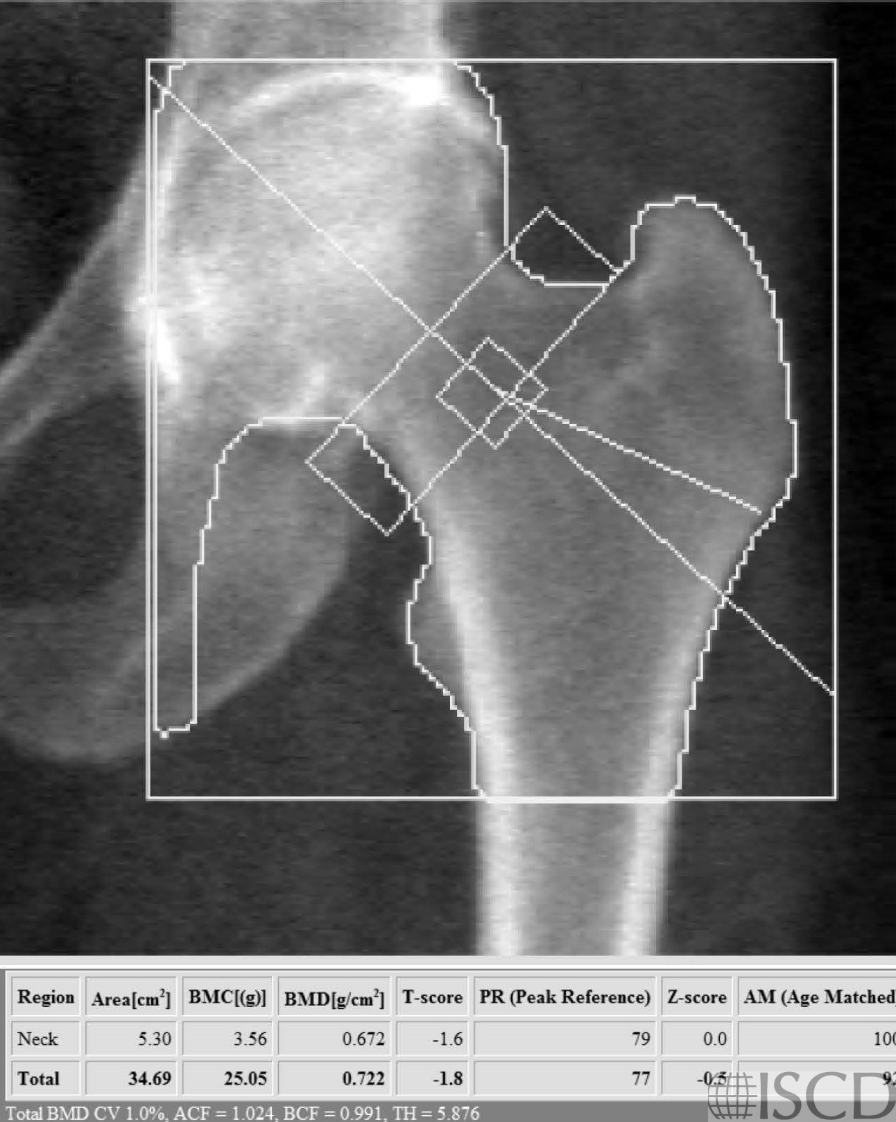
This is the left proximal femur Hologic scan from the same patient with hypermobile Ehlers-Danlos syndrome.
This is a DXA scan from a patient with hypermobile Ehlers-Danlos syndrome. The thoracic and lumbar spine radiographs showed demineralization and no other abnormalities. Bone mineral density can be variable in Ehlers-Danlos syndrome. Low bone mass is found in kyphoscoliotic, arthorchalasia (congenital hip dislocation, generalized joint hypermobility with recurrent joint dislocations), spondylodysplastic, and classic Ehlers-Danlos. In hypermobile and classic Ehlers-Danlos there are reports of mildly decreased bone density, but fracture risk may not always be increased.
Sarah L Morgan, MD, RD, CCD, The University of Alabama at Birmingham
• Basalom, S. and F. Rauch, Bone Disease in Patients with Ehlers-Danlos Syndromes. Curr Osteoporos Rep, 2020. 18(2): p. 95-102.
• Eller-Vainicher, C., et al., Bone involvement in adult patients affected with Ehlers-Danlos syndrome. Osteoporos Int, 2016. 27(8): p. 2525-31.
• Theodorou, S., D. Theodorou, and J. Adams, Bone loss in the axial and appendicular skeleton in Ehlers-Danlos Syndrome J Clin Densitom, 2012 15 p. S493