Hypophosphatasia

This is the Hologic lumbar spine scan from a postmenopausal female with hypophosphatasia.

This is the Hologic left proximal hip scan from a postmenopausal female with hypophosphatasia.
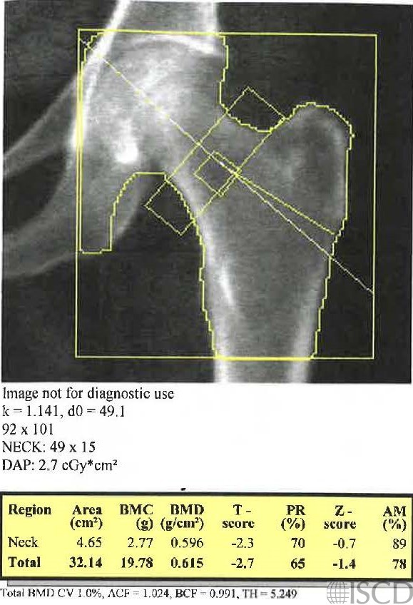
This DXA scan is from an 80-year-old Caucasian female who presented with low bone mass and a low alkaline phosphatase 20 U/L – normal low for lab = 37 U/L). A secondary cause workup showed an elevated pyridoxal phosphate level off of dietary supplements, and she had the diagnosis of hypophosphatasia. There is a compression fracture at L1, and L1 is omitted from the spine analysis. Other causes of a low alkaline phosphatase level include malnutrition, Wilson’s disease, hypothyroidism, zinc deficiency, vitamin C deficiency, hypophosphatemia, and pernicious anemia.
Sarah L Morgan, MD, RD, CCD, The University of Alabama at Birmingham
• Lefever, E., et al., Hypophosphatasia in Adults: Clinical Spectrum and Its Association With Genetics and Metabolic Substrates. J Clin Densitom, 2018.
• Simon, S., et al., Hypophosphatasia: From Diagnosis to Treatment. Curr Rheumatol Rep, 2018. 20(11): p. 69.
• Whyte, M.P., Hypophosphatasia – aetiology, nosology, pathogenesis, diagnosis and treatment. Nat Rev Endocrinol, 2016. 12(4): p. 233-46