Benign Bone Lesion – Enchondroma

This Hologic left proximal femur scan shows an enchondroma in the greater trochanter.
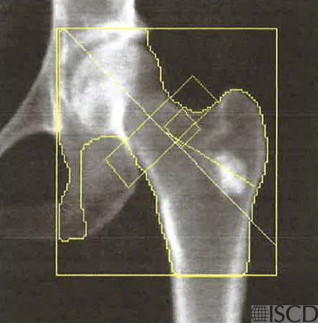
The companion radiograph shows the same lesion.

The DXA image shows an enchondroma in the greater trochanter. The accompanying radiograph shows the same lesion. The right proximal femur should be scanned and reported.
Sarah L Morgan, MD, RD, CCD, The University of Alabama at Birmingham
• Martineau, P., S. Bazarjani, and L.S. Zuckier, Artifacts and Incidental Findings Encountered on Dual-Energy X-Ray Absorptiometry: Atlas and Analysis. Semin Nucl Med, 2015. 45(5): p. 458-69.
• Kim, S.K. and W.F. Barry, Jr., Bone islands. Radiology, 1968. 90(1): p. 77-8.
• Geniets, C., et al., Proceedings of the European Society of Musculoskeletal Radiology (ESSR) training module, Antwerp, 20-21.01.05. Part two: bone tumors. Benign bone lesions: characteristic imaging features. Jbr-btr, 2006. 89(5): p. 266-74.