Cholelithiasis/Gallstones

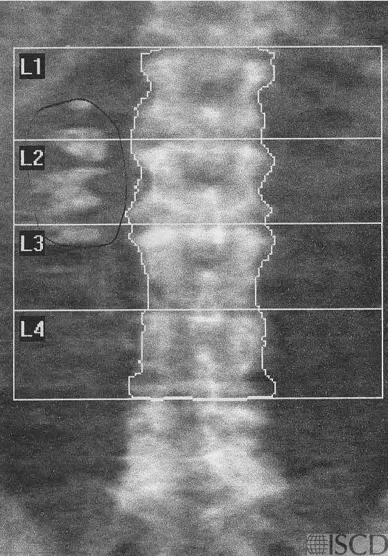
There are gallstones lateral to L1-L3 on the Hologic spine DXA scan.
The accompanying radiograph demonstrates the gallstones.
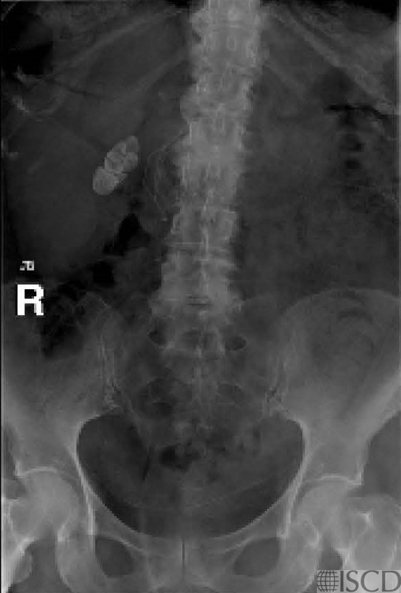
There are gallstones seen lateral to the Hologic spine on the right. The accompanying radiograph demonstrates the gallstones. The “undo” view may be checked in Hologic to confirm that the artifact is removed from the soft tissue baseline.
Sarah L Morgan, MD, RD, CCD, The University of Alabama at Birmingham
• Morgan, S.L., et al., The effect of common artifacts lateral to the spine on bone mineral density in the lumbar spine. J Clin Densitom, 2008. 11(2): p. 243-9.’ 6. Martineau, P., S. Bazarjani, and L.S. Zuckier, Artifacts and Incidental Findings Encountered on Dual-Energy X-Ray Absorptiometry: Atlas and Analysis. Semin Nucl Med, 2015. 45(5): p. 458-69.
• Bazzocchi, A., et al., Incidental findings with dual-energy X-ray absorptiometry: spectrum of possible diagnoses. Calcif Tissue Int, 2012. 91(2): p. 149-56.
• Martineau, P., S. Bazarjani, and L.S. Zuckier, Artifacts and Incidental Findings Encountered on Dual-Energy X-Ray Absorptiometry: Atlas and Analysis. Semin Nucl Med, 2015. 45(5): p. 458-69.
• Smith, J.A., R.P. Spencer, and D.P. Szigeti, Gall stones detected on lumbar bone densitometry examination. J Clin Densitom, 1998. 1(4): p. 403-4.