Gallstone

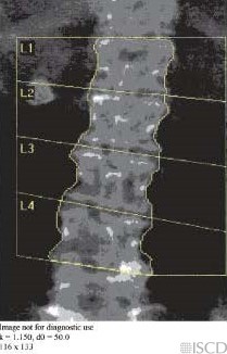
There is a gallstone lateral to L1-L2 on the right in this Hologic spine image. The undo view may be evaluated, to demonstrate that the gallstone is removed from the soft tissue baseline.
The accompanying radiograph shows the gallstone (blue circle).
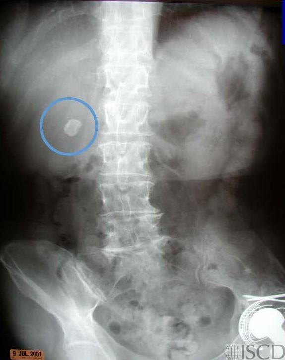
There is a gallstone lateral to L1-L2 on the Hologic lumbar spine scan. The accompanying radiograph shows the gallstone (blue circle).
E. Michael Lewiecki, MD, FACP, FACE, CCD, New Mexico Clinical Research & Osteoporosis Center
• Martineau, P., S. Bazarjani, and L.S. Zuckier, Artifacts and Incidental Findings Encountered on Dual-Energy X-Ray Absorptiometry: Atlas and Analysis. Semin Nucl Med, 2015. 45(5): p. 458-69.
• Bazzocchi, A., et al., Incidental findings with dual-energy X-ray absorptiometry: spectrum of possible diagnoses. Calcif Tissue Int, 2012. 91(2): p. 149-56.
• Bojinca, V., D. Opris, and M. Bonjinca, Artifacts and pitfalls in DXA scan images and interpretation. J Clin Densitom, 2012. 15(4): p. 486-487.