Klinefelter Syndrome

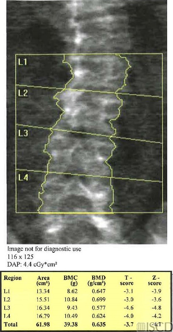
Hologic lumbar spine scan in a patient with Klinefelter Syndrome.
Hologic left proximal femur scan in a patient with Klinefleter Syndrome.
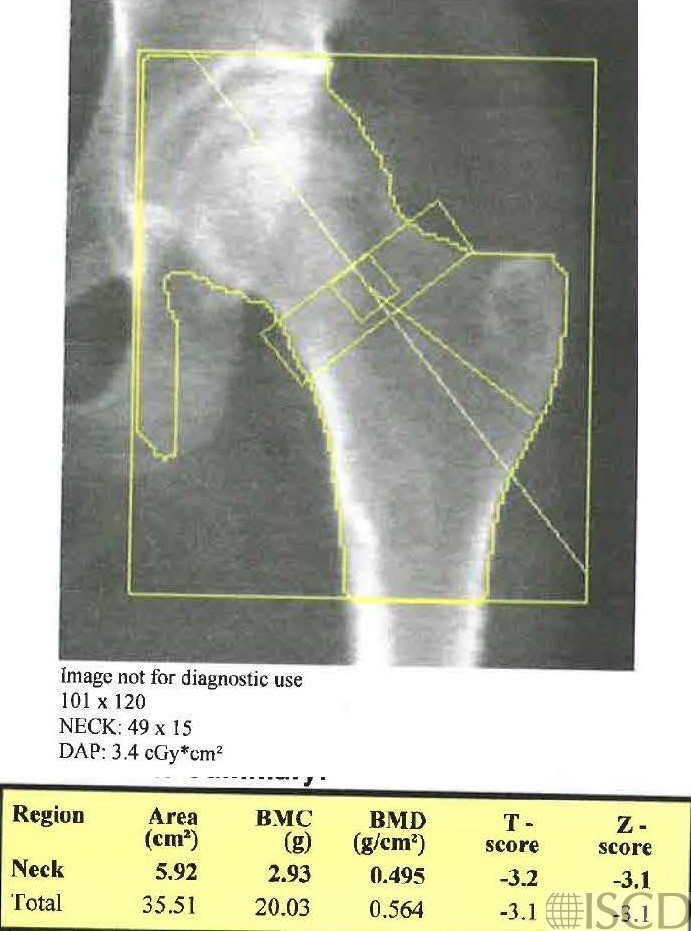
Klinefelter Syndrome is a random genetic error after conception, where a male is born with an extra X chromosome (XXY). Males have low testosterone levels and reduced muscle mass and low bone mineral density. There are degenerative changes in the lumbar spine which will overestimate the bone mineral density in this location.
Sarah L Morgan, MD, RD, CCD, The University of Alabama at Birmingham
• Orlic, Z.C. and L.G. Raisz, Causes of secondary osteoporosis. J Clin Densitom, 1999. 2(1): p. 79-92.
• Vieira da Costa, J., J.F. Pereira-Lima, and M. da Costa Oliveira, Bone mineral density in early-onset hypogonadism and the effect of hormonal replacement. J Clin Densitom, 2004. 7(3): p. 334-40.
A Ferlin 1, M Schipilliti, C Foresta. Bone density and risk of osteoporosis in Klinefelter syndrome. Acta Paediat 2011 Jun;100(6):878-84. doi: 10.1111/j.1651-2227.2010.02138.x. Epub 2011 Feb 10.
Piot A, Plotton I, Boutroy S, Bacchetta J, Ailloud S, Lejeune H, Chapurlat RD, Szulc P, Confavreux CB. Klinefelter Bone Microarchitecture Evolution with Testosterone Replacement Therapy. Calcif Tissue Int. 2022 Feb 13. doi: 10.1007/s00223-022-00956-2. Epub ahead of print. PMID: 35152305.
.
.