Biliary Drainage Catheter

This Hologic lumbar spine images shows a biliary drain tube lateral to the verebral bodies on the right.

The undo view shows that the artifact in the soft tissue is mapped and therefore removed from the soft tissue baseline.
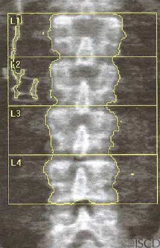
A biliary drainage catether is seen on the Hologic lumbar spine scan lateral to the vertebral bodies on the right. The undo view is also shown which documents that the artfiacts are mapped in the soft tissue and deleted from the soft tissue baseline. If an artifact overlies bone, the convention is to remove that spinal level. In Hologic, the undo view can be checked to confirm that the artifact is removed from the soft tissue baseline.
Sarah L Morgan, MD, RD, CCD, The University of Alabama at Birmingham.