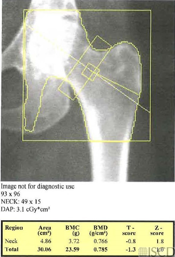
Femoral Neck Box Includes Part of the Ischium

In this image, the femoral neck box includes a portion of the ischium.

In this image, the ischium is deleted so that the femoral neck box data may be used. The length of the femoral neck box has now been changed to the default of 49 pixels.
On the analysis on the left, there is a portion of the ischium in the femoral neck box. The correct analysis is to paint out the ischium so that there is no ischium in the femoral neck box region of interest. The analysis on the right paints out the ischium so that the femoral neck box can be used and the femoral neck BMD is lower. There are many ways to paint out the ischium and drawing a straight line across is probably the most reproducible method of omitting the ischium. In the example on the right, a portion of the ischium is carved out, which would be difficult to reproduce on future scans.
Sarah L Morgan, MD, RD, CCD, The University of Alabama at Birmingham
• Feit, A., et al., Effect of positioning of the region of interest on bone density of the hip. . J Clin Densitom, 2020: 23(3) p 426-431.
• Morgan, S.L. and F. Peace, Do changes in the femoral neck box size make a significant difference in femoral neck BMD? . J Clin Densitom, 2011. 14 p. 156
• McKiernan, F.E., et al., A long femur scan field does not alter proximal femur bone mineral density measurements by dual-energy X-ray absorptiometry. J Clin Densitom, 2011. 14(3): p. 354-8.
• Prater, G.L., et al., The effect of extending femur scan length on BMD results on the Hologic Discovery-W scanner. J Clin Densitom, 2014. 17(4): p. 518-21.
• Celik, O., et al., The effect of hip rotation on bone mineral density of the proximal femur measured by dual energy X-ray absorptiometry. Eklem Hastalik Cerrahisi, 2009. 20(2): p. 71-7.
• Cheng, X.G., et al., Effects of anteversion on femoral bone mineral density and geometry measured by dual energy X-ray absorptiometry: a cadaver study. Bone, 1997. 21(1): p. 113-7.
• Girard, M.S., et al., Measured femoral density by dual-energy X-ray absorptiometry as a function of rotation. Orthop Rev, 1994. 23(1): p. 38-40.
• Goh, J.C., S.L. Low, and K. Bose, Effect of femoral rotation on bone mineral density measurements with dual energy X-ray absorptiometry. Calcif Tissue Int, 1995. 57(5): p. 340-3.
• Lekamwasam, S. and R.S. Lenora, Effect of leg rotation on hip bone mineral density measurements. J Clin Densitom, 2003. 6(4): p. 331-6.
• Rosenthall, L., Range of change of measured BMD in the femoral neck and total hip with rotation in women. J Bone Miner Metab, 2004. 22(5): p. 496-9.
• Tang, H., S.M. Ren, and X.Z. Luo, [Effect of femoral rotation on hip bone mineral density measurement]. Zhongguo Yi Xue Ke Xue Yuan Xue Bao, 2003. 25(3): p. 267-70.
• Wilson, C.R., et al., The effect of positioning on dual energy X-ray bone densitometry of the proximal femur. Bone Miner, 1991. 13(1): p. 69-76.
• McKiernan, F. and W. Washington, Effect of subtle positioning flaws on measured bone mineral density of the hip. J Clin Densitom, 2005. 8(3): p. 330-4.
• Hans, D., et al., Effects of a new positioner on the precision of hip bone mineral density measurements. J Bone Miner Res, 1997. 12(8): p. 1289-94.