GE-Lunar Short Femoral Neck ROI Placement Challenges

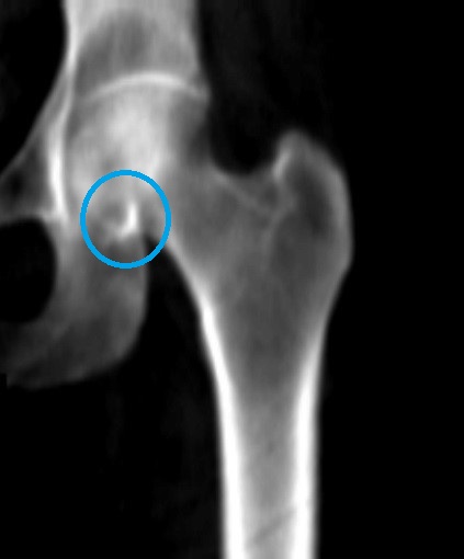
Keep the femoral neck ROI box from overlying the brighter whiter spot in the pelvis near the bottom of the femoral head identified by the blue circle in this image.
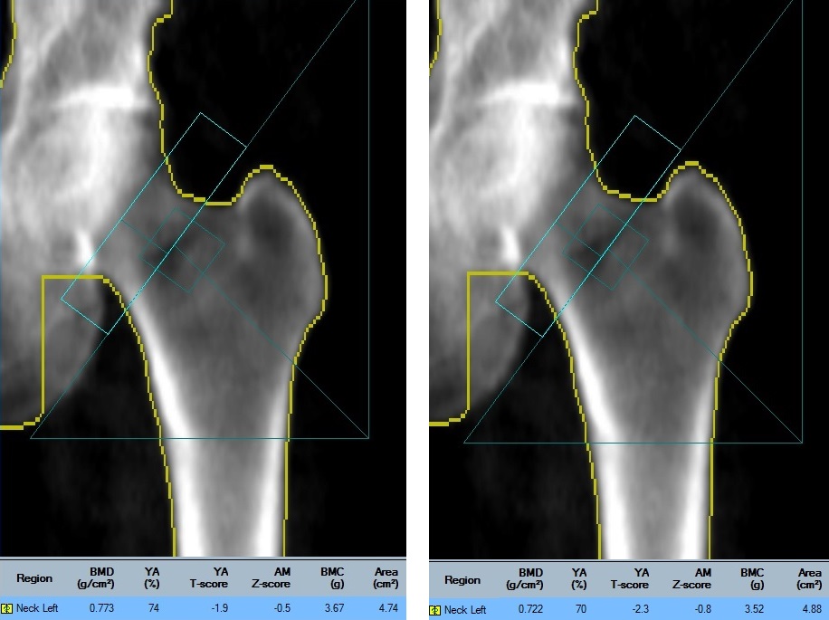
Two different placements of the neck rectangle for the same scan are shown here. When placed over the denser area of the pelvis, the ROI box calculates a falsely elevated the BMD. Excluding the pelvis from the analysis calculates the correct T-score of -2.3.
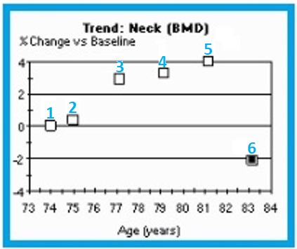
The numbered points on the graph correspond to the 6 numbered femur scans in image 2b. Notice that the three highest values are numbered 3, 4, 5. The three lowest are numbers 1, 2, 6.
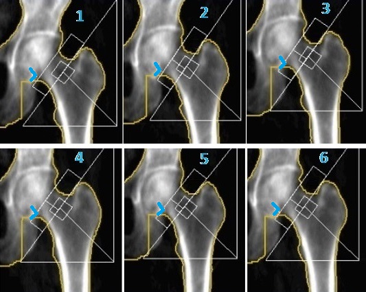
In femur scans 3, 4, and 5, the femoral neck rectangle overlies the brighter whiter bone of the pelvis, which is highlighted in blue here. In scans 1, 2, and 6, the neck box is positioned so that it excludes the denser bone that is not part of the femoral neck.
When a patient has a short femoral neck, the DXA technologist must use caution in placing the femur neck region-of-interest (ROI) rectangle to ensure that it does not overlie the denser area where the pelvis intersects the femur just below the femoral head.
If the neck box includes the denser pelvis bone, the BMD will be falsely elevated. This is something that should be considered when interpreting serial scans. Out of the six numbered femur scans, the most reliable measurements are numbers 1, 2, and 6. When scan 6 is compared to scan 5, there appears to have been a BMD loss that exceeds the LSC for this facility, however this is likely incorrect. Scan 6 is more appropriately compared to numbers 1 and 2 from which there has been no significant change.
NOTE: These are GE-Lunar scans. The femoral neck ROI box placement differs from that of other DXA manufacturers.
Wendy Tolman-Andrews, BS, RT(BD), CBDT, UConn Health, Radiology, Musculoskeletal Institute, Farmington, CT