Uterine Fibroid

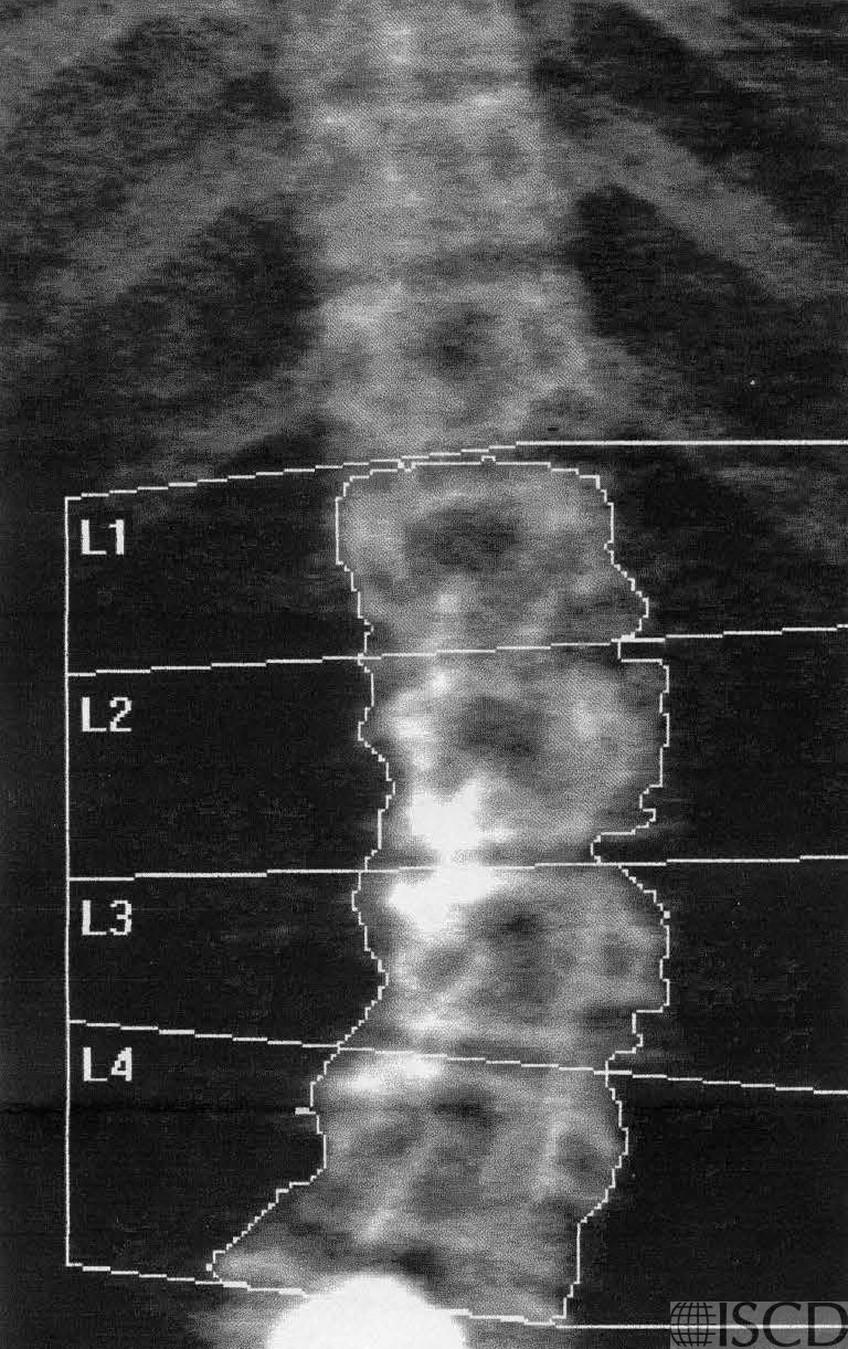
There is a calcified fibroid partially overlying L4 on this lumbar spine DXA scan.
A calcified uterine fibroid is seen within the pelvic brim. The pelvic ultrasound showed a 6.2 x 4.1 x 6.4 cm uterine fibroid int he left mid uterine body. The fibroid is not within the region of interest scan in the left proximal DXA scan.
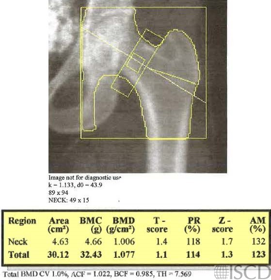
The DXA scans show examples of uterine fibroids seen as internal artifacts.
Laura Carbone, MD, CCD, Augusta University Medical Center and Charlie Norwood Veterans Affairs Medical Center (top image)
Sarah L Morgan, MD, RD, CCD, The University of Alabama at Birmingham (bottom image)
• Martineau, P., S. Bazarjani, and L.S. Zuckier, Artifacts and Incidental Findings Encountered on Dual-Energy X-Ray Absorptiometry: Atlas and Analysis. Semin Nucl Med, 2015. 45(5): p. 458-69.
• Morgan, S.L. and G.L. Prater, Quality in dual-energy X-ray absorptiometry scans. Bone 2017 Nov; 104:13-28.
• Bazzocchi, A., et al., Incidental findings with dual-energy X-ray absorptiometry: spectrum of possible diagnoses. Calcif Tissue Int, 2012. 91(2): p. 149-56.