Calcified Tendon

There is a calciified tendon superior to the left greater trochanter seen on the left Hologic proximal femur image.
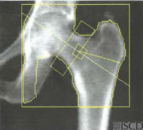
The calcified area at the top of the greater trochanter represents a calcified tendon. The calcified tendon is not outlined in the bone map of the left proximal femur.
Sarah L Morgan, MD, RD, CCD, The University of Alabama at Birmingham
• Bazzocchi, A., et al., Incidental findings with dual-energy X-ray absorptiometry: spectrum of possible diagnoses. Calcif Tissue Int, 2012. 91(2): p. 149-56.