Pacemaker

DXA report

Chest X-ray
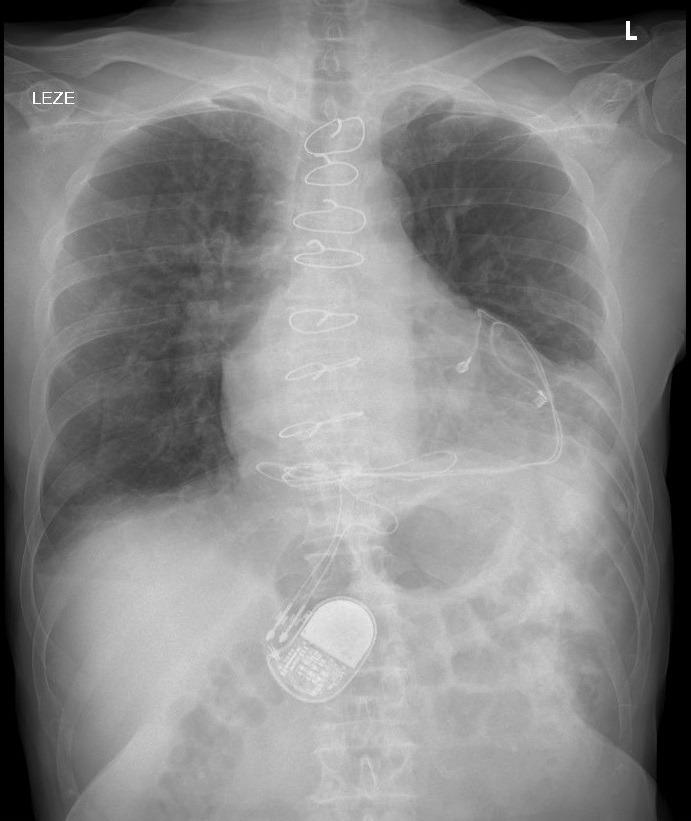
This lumbar spine DXA scan shows an artifact over the L1 vertebral body. After careful assessment of the DXA image, a pacemaker was identified and confirmed with evaluation of the prior chest x-ray images. BMD was measured in a male patient, as part of comprehensive follow-up for osteoporosis and endocrine abnormalities in post-heart transplant patients. L1 was omitted because of the overlying artifact. L4 was omitted because of degenerative change and discordance.
External or internal artifacts, such as bra wires, body jewelry, spinal fusion hardware, radiopaque medications are commonly encountered in DXA images. Because pacemakers are usually placed under collarbone, they are not seen on DXA image. However, in adults with heart surgery and children, often pacemakers with epicardial lead placement are used. Epicardial pacemakers are inserted in retrocostal, subxiphoid or subrectus location and can distort DXA measurements in this specific population.
Matej Rakusa, MD, PhD, CCD, Department of Endocrinology, Diabetes and Metabolic Disease, University Medical Centre Ljubljana ,Faculty of Medicine, University of Ljubljana.
Martineau P, Bazarjani S, Zuckier LS. Artifacts and Incidental Findings Encountered on Dual-Energy X-Ray Absorptiometry: Atlas and Analysis. Semin Nucl Med. 2015 Sep;45(5):458-69.
Lichtenstein BJ, Bichell DP, Connolly DM, Lamberti JJ, Shepard SM, Seslar SP. Surgical approaches to epicardial pacemaker placement: does pocket location affect lead survival?. Pediatr Cardiol. 2010;31(7):1016-1024. doi:10.1007/s00246-010-9754-1