Metastatic Breast Cancer

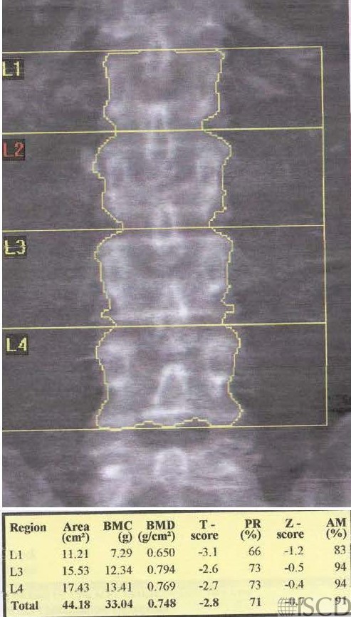
This is a baseline Hologic lumbar spine DXA scan.
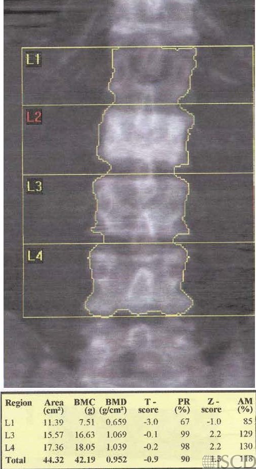
This is a Hologic DXA scan 4 years later showing sclerosis at L2. There are also large increases in bone mineral density at L3 and L4 since the baseline scan.
This is the accompanying scout film from a CT scan. L2, L3, and L4 also look sclerotic.

This is the nuclear medicine scan. The biopsy- proven diagnosis was metastatic breast cancer.
This case shows a DXA scan, with a follow-up scan 4 years later showing a sclerotic L2 and large increases in bone mineral density. This ultimately was proven to be metastatic breast cancer. The accompanying radiograph and nuclear medicine scan are also included.
Sarah L Morgan, MD, RD, CCD, The University of Alabama at Birmingham
• Fraenkel, M., et al., Association between bone mineral density and incidence of breast cancer. PLoS One, 2013. 8(8): p. e70980.
• Kim, B.K., et al., Bone mineral density and the risk of breast cancer: a case-control study of Korean women. Ann Epidemiol, 2014. 24(3): p. 222-7.
• Gupta, R., et al., Relationship between mammography breast density and bone mineral density. J Clin Densitom, 2008. 11(3): p. 431-6.