Pelvic Coils

This image shows the pelvic coils lateral to L2-L4 on the Hologic spine image. The pelvic coils show up as a black hole-type artifact.
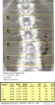
Because the coils overlapped L3 and L4, these levels were initially removed from analysis.
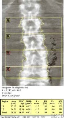
This image shows the undo view for the lumbar spine image. Notice that the coils are not removed from the soft tissue baseline, this means that the presence of the coils will affect the accuracy of measurement of L1-L2.

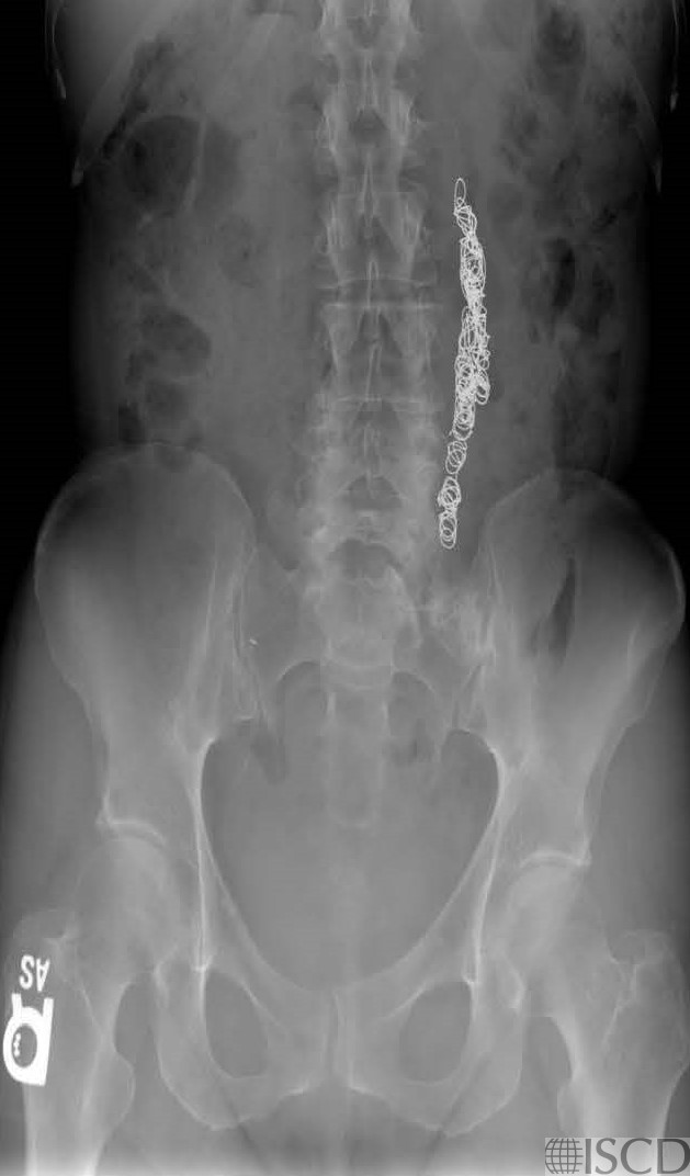
This is the accompanying abdominal radiograph showing the pelvic coils.
Pelvic coils are seen lateral to L2-L4 on the Hologic lumbar spine image. Pelvic coils are used for pelvic congestion syndrome. They are said to relieve pain by closing off faulty vein so they can’t become enlarged with blood. The coils show up as black hole-type artifacts.
Sarah L Morgan, MD, RD, CCD, The University of Alabama at Birmingham
• Guirola, J.A., et al., A Randomized Trial of Endovascular Embolization Treatment in Pelvic Congestion Syndrome: Fibered Platinum Coils versus Vascular Plugs with 1-Year Clinical Outcomes. J Vasc Interv Radiol, 2018. 29(1): p. 45-53.
• Lopez, A.J., Female Pelvic Vein Embolization: Indications, Techniques, and Outcomes. Cardiovasc Intervent Radiol, 2015. 38(4): p. 806-20.