Hip Osteoarthritis – How Bad Can It Get?

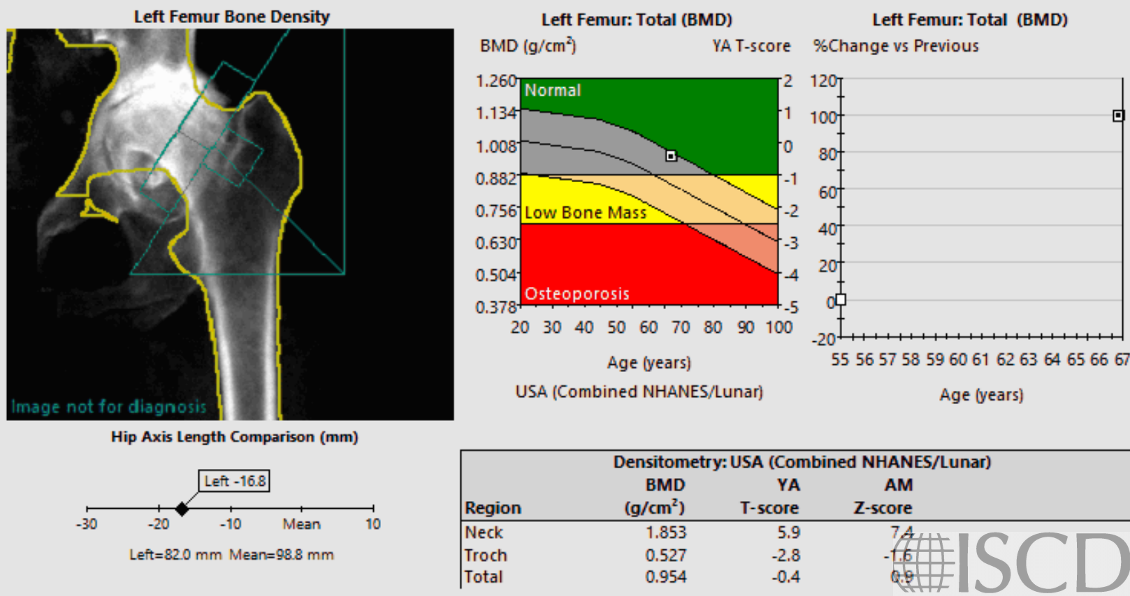
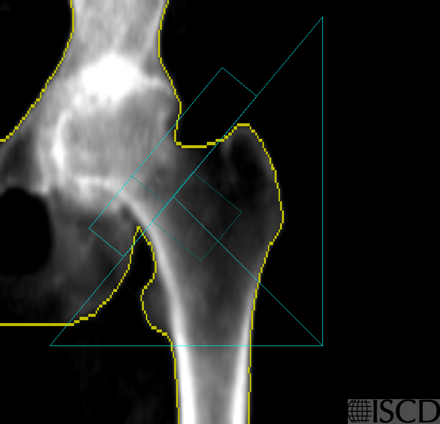
Left hip DXA shows marked site discordance, from +5.9 to the femur neck to -2.8 for the trochanter. Dense sclerosis is seen to affect the femur neck ROI with advanced OA and erosive loss of the femoral head. (Note that it is impossible to achieve adequate hip positioning with soft tissue on both sides of the neck.)
Left hip DXA from 12 years earlier showed osteoporosis (total hip T-score -4.2) with milder effects from osteoarthritis (femur neck T-score -2.2) .
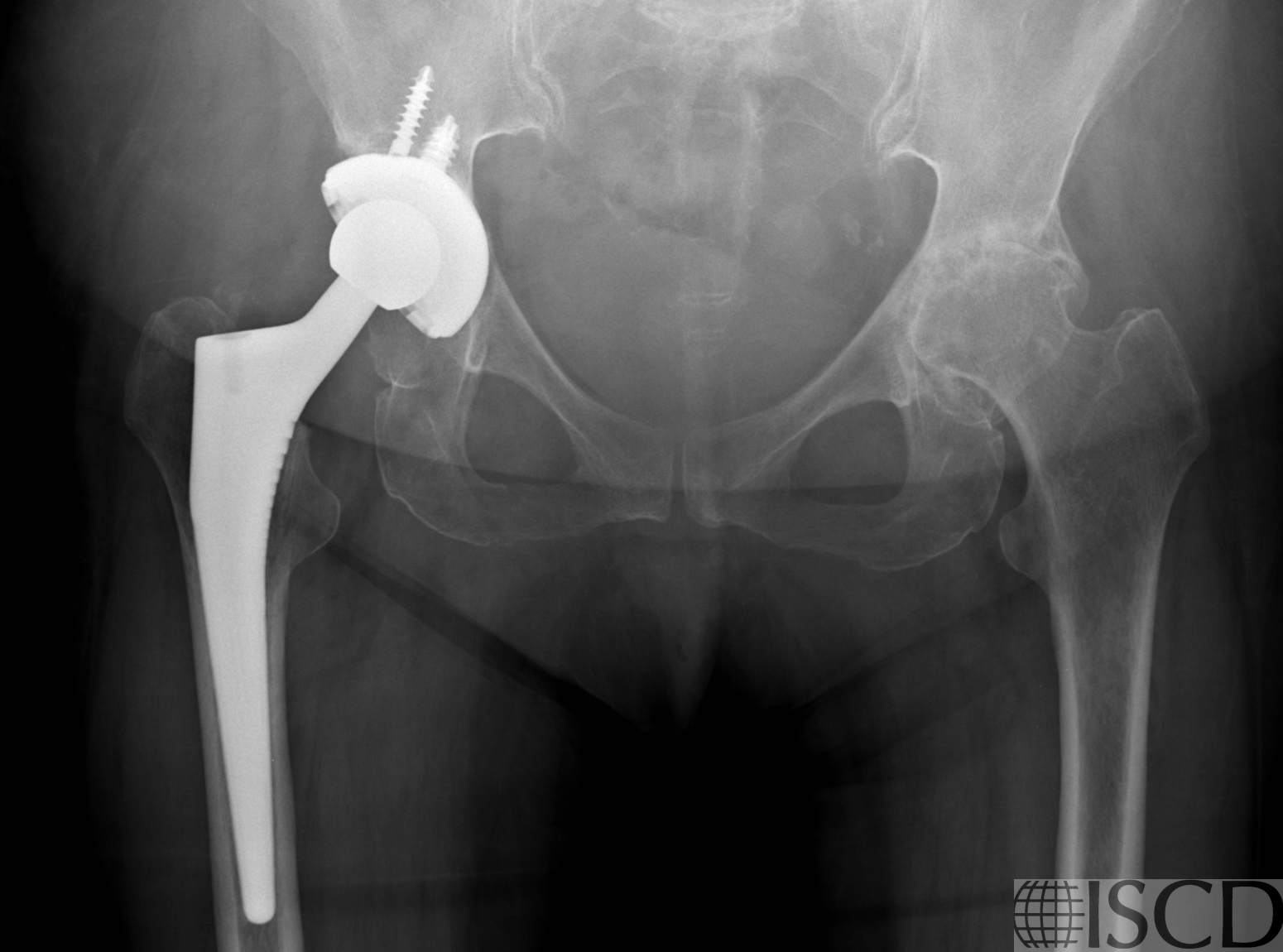
X-ray from 10 years earlier show bone-on-bone osteoarthritis affecting the left hip, previous right arthroplasty.
Forearm one-third radius T-score -3.8 (osteoporotic).

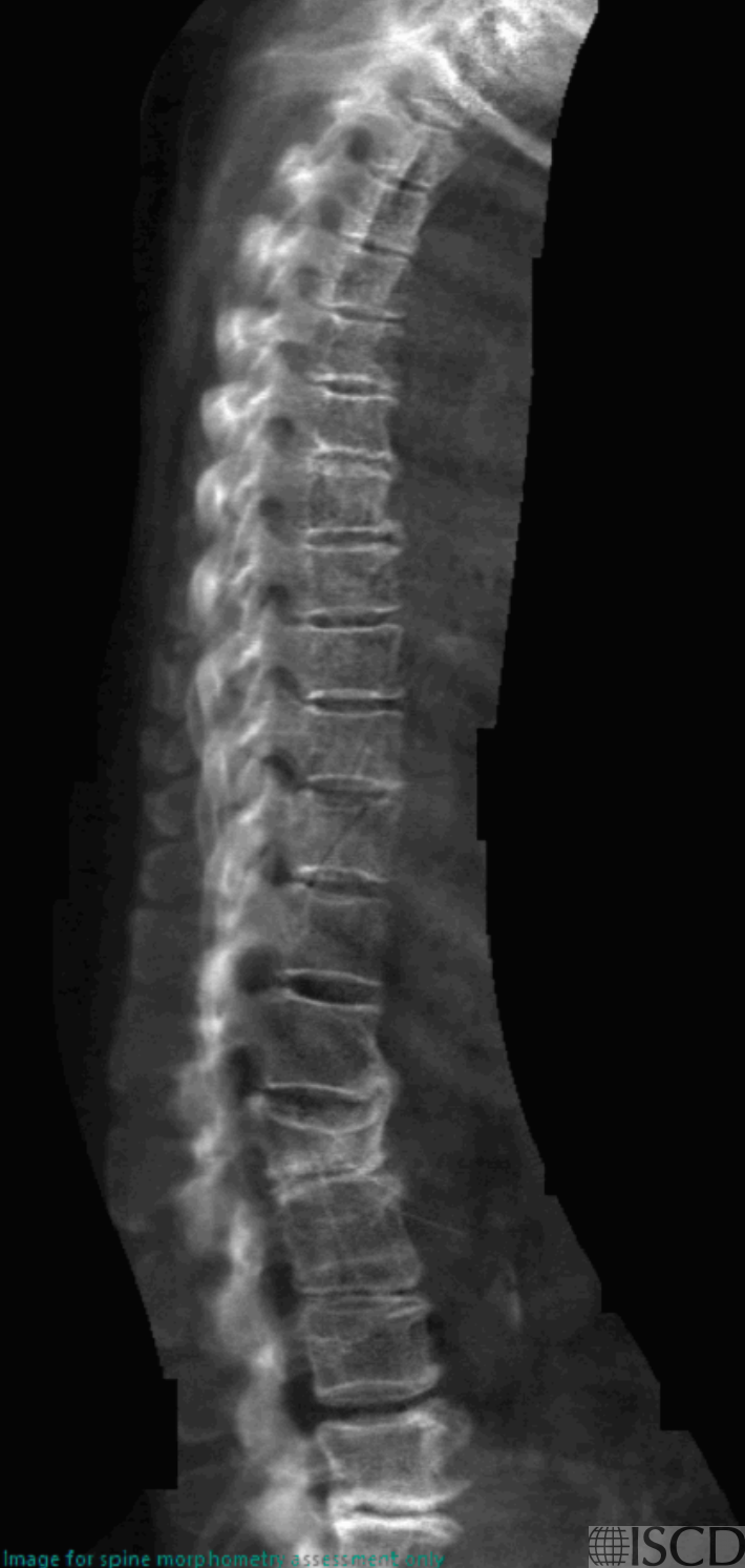
VFA shows a severe L2 compression fracture, new since 2010.
66 year old woman with DXA assessment prior to starting aromatase inhibitor therapy. The spine was not usable due to degenerative changes and intercurrent compression fracture (see VFA). The hip is less often affected by degenerative changes but osteoarthritis can produce overestimation in BMD. The femur neck is usually most severely affected while the trochanter (not a standard reporting site) is least affected.
Left femur neck BMD was 0.731 g/cm2 in 2010 (indicating an interval increase of 1.122 g/cm2!!). Left total hip BMD was 0.569 g/cm2 in 2010 (indicating an increase of 0.385 g/cm2). Although she had received oral BP therapy 2011-2016, this cannot produce such a dramatic increase.
Note that she meets two definitions of osteoporosis — forearm one-third T-score below -2.5, and also a low-trauma vertebral fracture.
William D. Leslie, MD FRCPC MSc CCD, University of Manitoba