Acetabula Protrusio

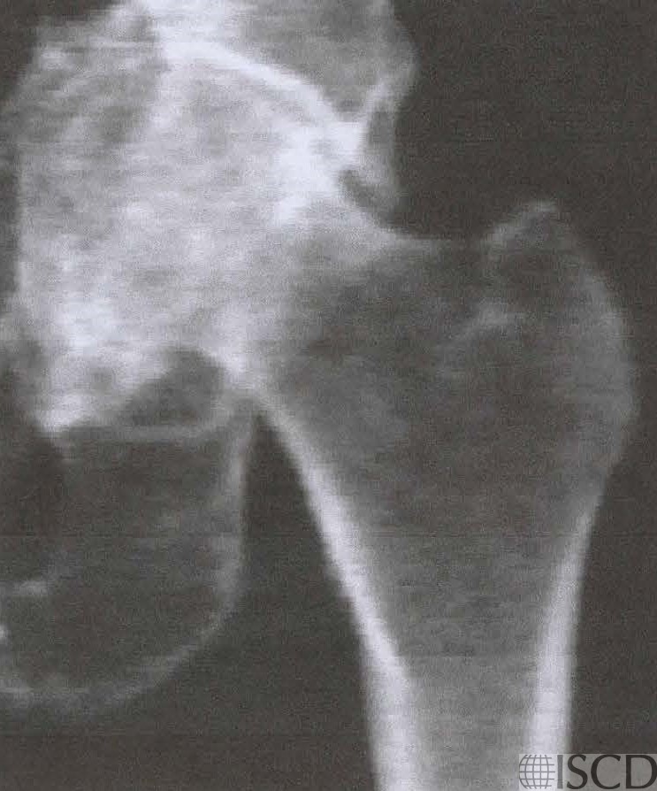
The Hologic DXA image of the left hip shows acetabula protrusio.
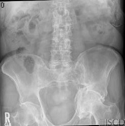
The accompanying radiograph shows the left acetabula protrusio.
This is a patient with acetabular protrusion. The right hip would need to be used for the DXA scan. Protrusio acetabuli can be primary and is often associated with osteoarthritis. It may also be familial. Secondary causes include Paget’s disease, psoriatic arthropathy, rheumatoid arthritis, ankylosing spondylitis, osteomalacia and rickets, osteogenesis imperfecta, Marfan syndrome, trauma, and hemophilia.
Sarah L Morgan, MD, RD, CCD, The University of Alabama at Birmingham
• Ahn, J., et al., Acetabular Protrusio in Patients With Osteogenesis Imperfecta: Risk Factors and Progression. J Pediatr Orthop, 2017.
• De Maio, F., et al., Orthopaedic Aspects of Marfan Syndrome: The Experience of a Referral Center for Diagnosis of Rare Diseases. Adv Orthop, 2016. 2016: p. 8275391.
• Saglam, Y., et al., Total hip arthroplasty in patients with ankylosing spondylitis: Midterm radiologic and functional results. Acta Orthop Traumatol Turc, 2016. 50(4): p. 443-7.
• Dutka, J., et al., Total hip arthroplasty with bone grafts for protrusio acetabuli. Ortop Traumatol Rehabil, 2011. 13(5): p. 469-77.
• Fitzpatrick, L.A., Secondary causes of osteoporosis. Mayo Clin Proc, 2002. 77(5): p. 453-68.
• Kok, C. and P.N. Sambrook, Secondary osteoporosis in patients with an osteoporotic fracture. Best Pract Res Clin Rheumatol, 2009. 23(6): p. 769-79.
• Orlic, Z.C. and L.G. Raisz, Causes of secondary osteoporosis. J Clin Densitom, 1999. 2(1): p. 79-92.
• Stein, E. and E. Shane, Secondary osteoporosis. Endocrinol Metab Clin North Am, 2003. 32(1): p. 115-34, vii.