Benign Bone Lesion – Enchondroma

There is an enchondroma seen in the left femoral shaft on this Hologic left proximal femur radiograph.
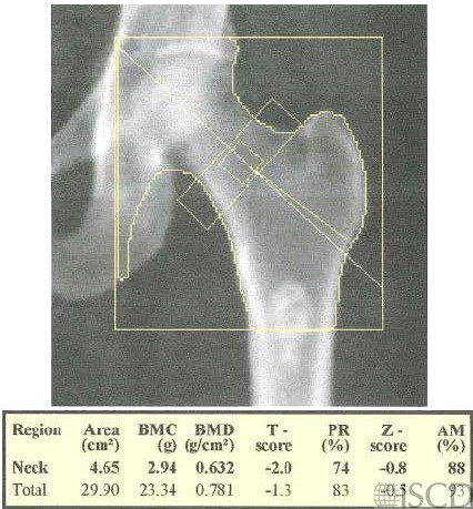
On the accompanying radiograph, the lesion was read as an enchondroma.

The DXA image shows a left Hologic proximal femur scan with an enchondroma in the left femoral shaft. The accompanying radiograph was read as an enchondroma. An enchondroma is a cartilage cyst found in the bone marrow. Enchondromas have a characteristic appearance on MRI scanning. An enchondroma may have increased uptake on a PET scan.
Sarah L Morgan, MD, RD, CCD, The University of Alabama at Birmingham
• Martineau, P., S. Bazarjani, and L.S. Zuckier, Artifacts and Incidental Findings Encountered on Dual-Energy X-Ray Absorptiometry: Atlas and Analysis. Semin Nucl Med, 2015. 45(5): p. 458-69.
• Kim, S.K. and W.F. Barry, Jr., Bone islands. Radiology, 1968. 90(1): p. 77-8.
• Geniets, C., et al., Proceedings of the European Society of Musculoskeletal Radiology (ESSR) training module, Antwerp, 20-21.01.05. Part two: bone tumors. Benign bone lesions: characteristic imaging features. Jbr-btr, 2006. 89(5): p. 266-74.