Primary Hyperparathyroidism

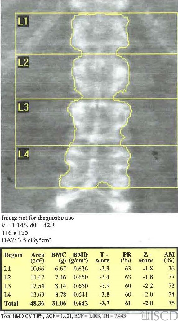
This is the Hologic spine scan from a patient with surgery-proven case primary hyperparathyroidism .
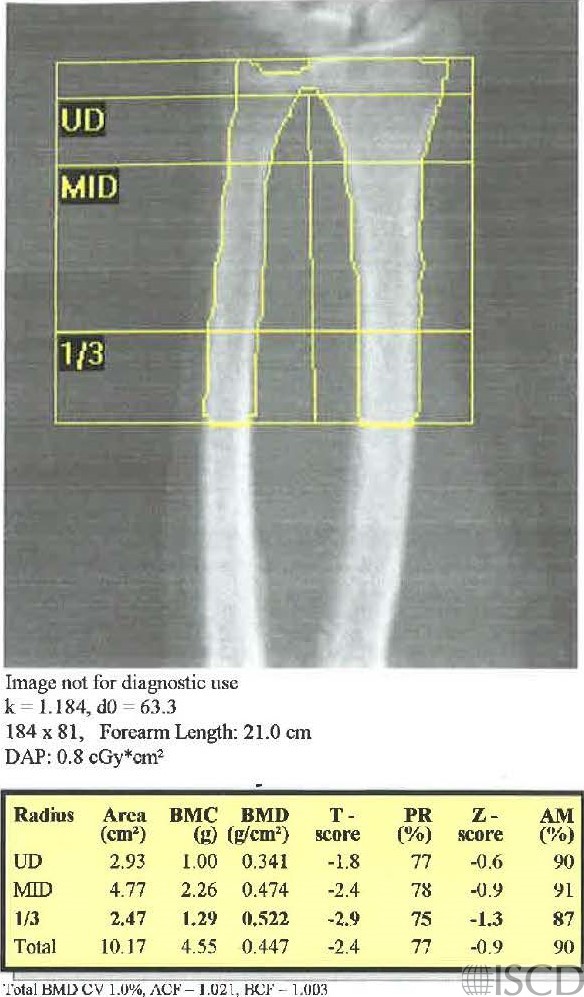
This is the Hologic left wrist scan from a surgery-proven case of primary hyperparathyroidism.
This is the Hologic left proximal hip scan from a surgery-proven case of primary hyperparathyroidism.
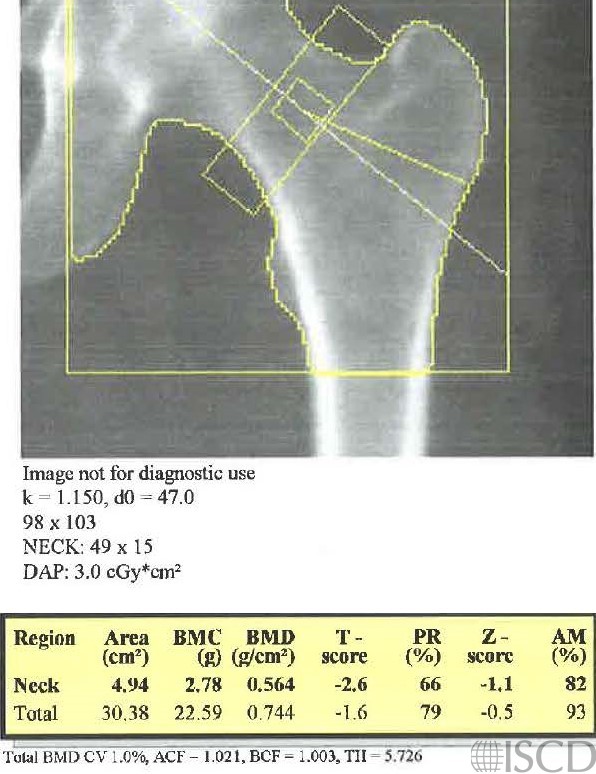
This is the baseline DXA scan from a postmenopausal woman referred to Osteoporosis Clinic for low bone density and hypercalcemia. The intact parathyroid hormone level was elevated. A parathyroid adenoma was resected. A year later she had recurrent hypercalcemia and elevation of the intact parathyroid hormone level, and an additional adenoma was found and resected. Completing a wrist scan in a patient with hyperparathyroidism is important as an assessment of cortical bone mineral density.
Sarah L Morgan, MD, RD, CCD, The University of Alabama at Birmingham
• Lewiecki, E.M. and P.D. Miller, Skeletal effects of primary hyperparathyroidism: bone mineral density and fracture risk. J Clin Densitom, 2013. 16(1): p. 28-32.
• Bilezikian, J.P., Primary Hyperparathyroidism. J Clin Endocrinol Metab, 2018. 103(11): p. 3993-4004.
• Miguel, G.A., et al., Trabecular Bone Score, Bone Mineral Density and Bone Markers in Patients with Primary Hyperparathyroidism 2 Years After Parathyroidectomy. Horm Metab Res, 2019. 51(3): p. 186-190.
• Castellano, E., et al., Forearm DXA Increases the Rate of Patients With Asymptomatic Primary Hyperparathyroidism Meeting Surgical Criteria. J Clin Endocrinol Metab, 2016. 101(7): p. 2728-32.
• Dutta, D., et al., Severity and pattern of bone mineral loss in endocrine causes of osteoporosis as compared to age-related bone mineral loss. J Postgrad Med, 2016. 62(3): p. 162-9.
• Silverberg, S.J., et al., Current issues in the presentation of asymptomatic primary hyperparathyroidism: proceedings of the Fourth International Workshop. J Clin Endocrinol Metab, 2014. 99(10): p. 3580-94