Hip Rotation

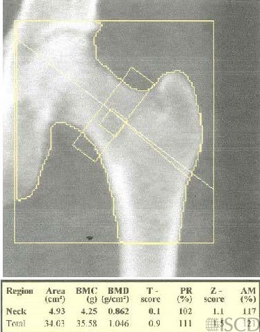
This Hologic hip scan has the femur straight and the hip is internally rotated.
This Hologic hip scan is on the same individual, on the same day with the hip externally rotated. The rotation of the hip is gauged by the amount of lesser trochanter that is seen. The bone mineral density of the total hip is not affected by the difference in rotation in this example.
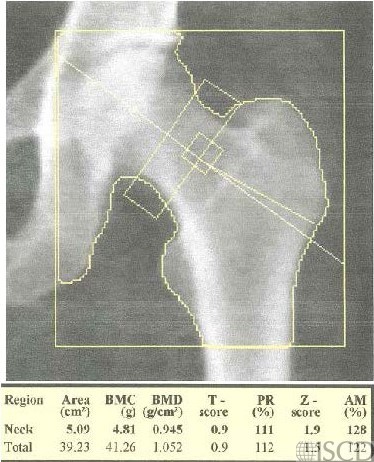
Differences in hip rotation will cause corresponding differences in the femoral neck bone mineral density. Differences in rotation of the hip can affect both the accuracy and precision of hip DXA measurements. The optimal positioning of the hip is to have the femoral neck parallel to the table, which means that the hip is internally rotated with the shaft of the femur straight. Numerous investigators have found that differences in hip rotation impact both precision and accuracy (see references below). McKiernan and Washington concluded that subtle differences in hip positioning on a GE/Lunar Prodigy scanner didn’t generate predictable changes in BMD at any of the hip regions of interest (reference below). The use of hip positioning devices can help with consistent hip positioning. There is a tiny amount of greater trochanter in this scan, this could easily be reanalyzed.
slmorgan@uab.edu
205-834-6441
• Celik, O., et al., The effect of hip rotation on bone mineral density of the proximal femur measured by dual energy X-ray absorptiometry. Eklem Hastalik Cerrahisi, 2009. 20(2): p. 71-7.
• Cheng, X.G., et al., Effects of anteversion on femoral bone mineral density and geometry measured by dual energy X-ray absorptiometry: a cadaver study. Bone, 1997. 21(1): p. 113-7.
• Girard, M.S., et al., Measured femoral density by dual-energy X-ray absorptiometry as a function of rotation. Orthop Rev, 1994. 23(1): p. 38-40.
• Goh, J.C., S.L. Low, and K. Bose, Effect of femoral rotation on bone mineral density measurements with dual energy X-ray absorptiometry. Calcif Tissue Int, 1995. 57(5): p. 340-3.
• Lekamwasam, S. and R.S. Lenora, Effect of leg rotation on hip bone mineral density measurements. J Clin Densitom, 2003. 6(4): p. 331-6.
• Rosenthall, L., Range of change of measured BMD in the femoral neck and total hip with rotation in women. J Bone Miner Metab, 2004. 22(5): p. 496-9.
• Tang, H., S.M. Ren, and X.Z. Luo, [Effect of femoral rotation on hip bone mineral density measurement]. Zhongguo Yi Xue Ke Xue Yuan Xue Bao, 2003. 25(3): p. 267-70.
• Wilson, C.R., et al., The effect of positioning on dual energy X-ray bone densitometry of the proximal femur. Bone Miner, 1991. 13(1): p. 69-76.
• McKiernan, F. and W. Washington, Effect of subtle positioning flaws on measured bone mineral density of the hip. J Clin Densitom, 2005. 8(3): p. 330-4.
• Hans, D., et al., Effects of a new positioner on the precision of hip bone mineral density measurements. J Bone Miner Res, 1997. 12(8): p. 1289-94.