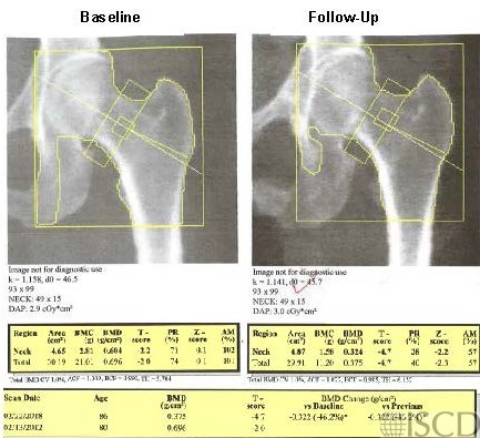
Follow-up Hip Scan with the Lesser Trochanter Incompletely Outlined


The left panel shows the baseline scan and the right panel shows the follow up scan with the lesser trochanter not outlined.

This panel shows the corrected scan with the ischium deleted in approximately the same way and the lesser trochanter is now outlined.

Case Description:

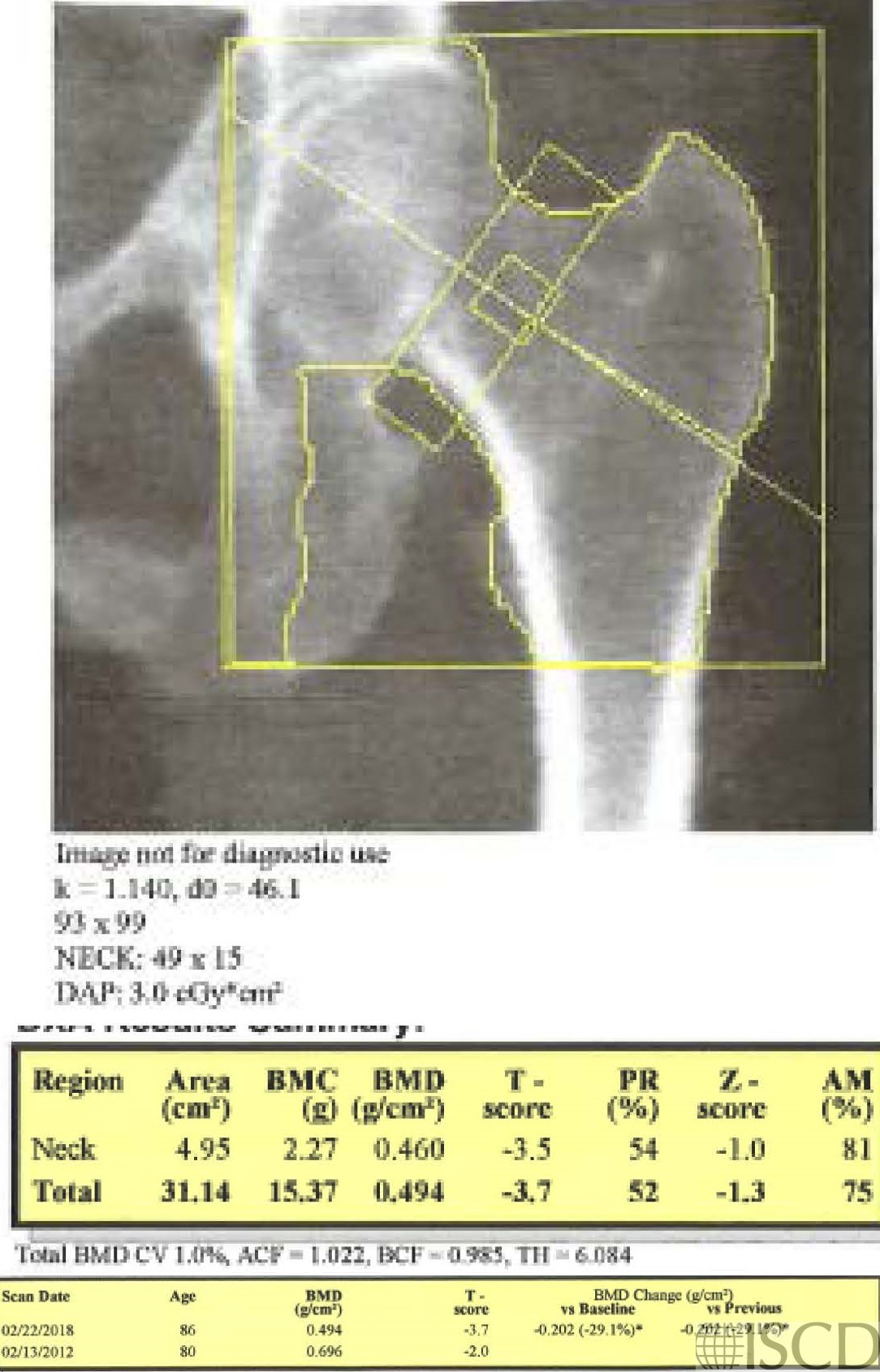
The baseline scan is presented in the upper left panel. In the upper right panel is the follow-up scan. The ischium was omitted differently, and the lesser trochanter was not outlined. The bottom panel shows the corrected scan with the ischium omitted in approximately the same way and the lesser trochanter now outlined. The total hip bone mineral density is higher with the lesser trochanter outlined. The scans were 6 years apart and there is a significant difference at the total hip with both scenarios, however the magnitude of the loss is much greater without the lesser trochanter being outlined.

Credit:

Sarah L Morgan, MD, RD, CCD, The University of Alabama at Birmingham

References:
  • Watts, N.B., Fundamentals and pitfalls of bone densitometry using dual-energy X-ray absorptiometry (DXA). Osteoporos Int, 2004. 15(11): p. 847-54.
  • Choplin R.H., Lenchik L and S. Wuertzer, A practical approach to interpretation of Dual-Energy X-ray Absorptiometry (DXA) for assessment of bone density. . Curr Radiol Rep  2(48).
  • Dasher, L.G., C.D. Newton, and L. Lenchik, Dual X-ray absorptiometry in today’s clinical practice. Radiol Clin North Am, 2010. 48(3): p. 541-60.
  • Theodorou, D.J. and S.J. Theodorou, Dual-energy X-ray absorptiometry in clinical practice: application and interpretation of scans beyond the numbers. Clin Imaging, 2002. 26(1): p. 43-9.
  • Mergler, S., et al., Lumbar spine and total-body dual-energy X-ray absorptiometry in children with severe neurological impairment and intellectual disability: a pilot study of artefacts and disrupting factors. Pediatr Radiol, 2012. 42(5): p. 574-83.
  • Choi, J.S., The influence of soft tissue recognition errors on BMD value-A case report: Recipient of Young Investigator Award J Clin Densitom, 2012. 15(4): p. 483
  • Fuleihan, G.E., et al., Reproducibility of DXA absorptiometry: a model for bone loss estimates. J Bone Miner Res, 1995. 10(7): p. 1004-14.
  • Fuerst, T., et al., Quality Assurance in Bone Densitometry in Bone Densitometry and Osteoporosis  K. Genant, G. Guglielmi, and M. Jergas, Editors. 1998, Springer Berlin.
  • Hansen, K., et al., DXA Errors are Common and Likely Adversely Affect Clinical Care: DXA Quality Improvement is Needed. J Bone Miner Res 2016. 31((Suppl 1) Available at http://www.asbmr.org/ItineraryBuilder/Presentationaspx?pid=83c01c31-237b-4f07-81a5-1eeb2a7968aa&ptag=AuthorDetail&aid=00000000-0000-0000-0000-000000000000. ).
  • Promma, S., et al., Errors in Patient Positioning for Bone Mineral Density Assessment by Dual x-ray Absorptiometry: Effect of Technologist Retraining. J Clin Densitom, 2018. 21(2): p. 252-259.
  • Cetin, A., et al., Evaluation of the patient positioning during DXA measurements in daily clinical practice. Clin Rheumatol, 2008. 27(6): p. 713-5.
  • Staron, R.B., et al., Computerized bone densitometric analysis: operator-dependent errors. Radiology, 1999. 211(2): p. 467-70.
  • Baniak, N., S. Grzybowski, and W.P. Olszynski, Dual-energy x-ray absorptiometry scan autoanalysis vs manual analysis. J Clin Densitom, 2014. 17(1): p. 97-103.