Bone Densitometry study in patient with metastatic hormone sensitive prostate cancer

Bone density analysis of the lumbar spine (L1 to L4)

Bone density analysis of the left forearm

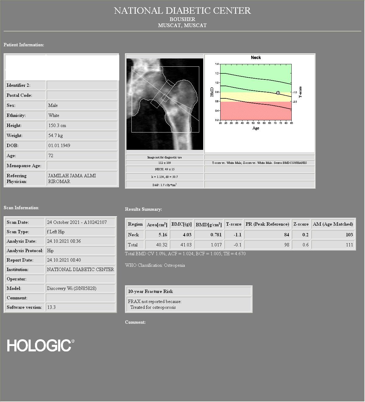
Bone density analysis of the left hip
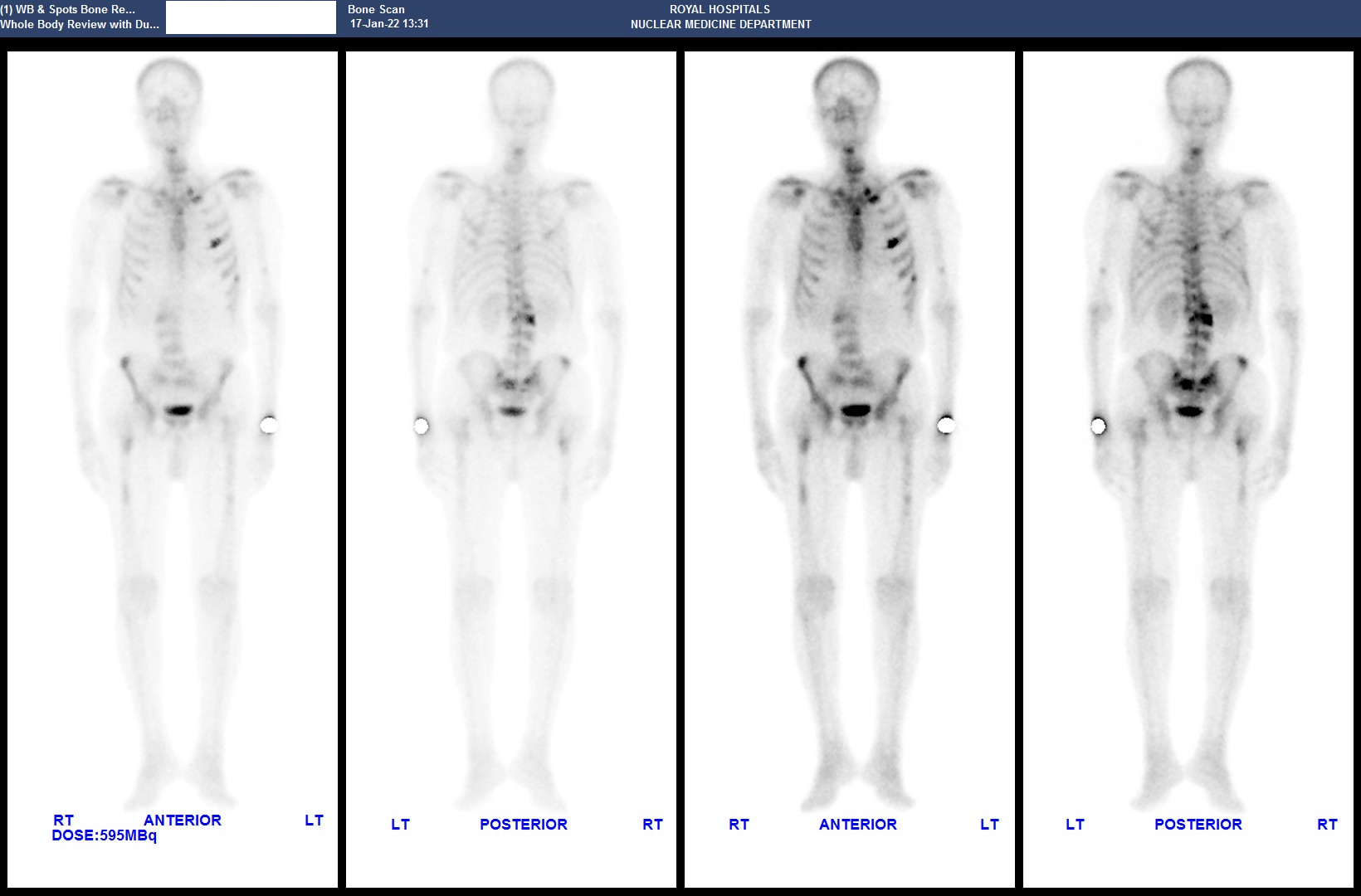
Whole body bone scintigraphy
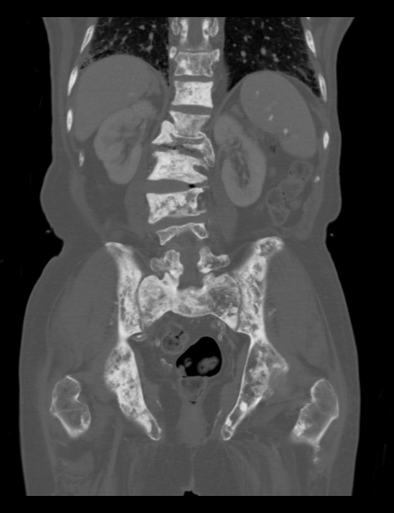
Abdomen and pelvic CT scan, coronal image in bone window
Abdomen and pelvis CT scan, sagittal image in bone window

72 years old male patient a newly diagnosed case of hormone sensitive metastatic prostate cancer. BMD study was done to evaluate for bone density status.
The study was done at our institution in Royal Hospital, Muscat, Oman.
The BMD machine used is Hologic, model: Discovery Wi (S/N85828)
The non diagnostic image for the Lumbar spine (L1 to L4) used for BMD analysis showed diffuse sclerotic changes involving all the lumbar spine vertebrae as well as distal thoracic spine vertebrae with collapse of L1 vertebral body.
The T-score of the Total spine was 2.7
The non diagnostic image of the left hip used for BMD analysis showed ill-defined rounded sclerotic lesions in the femoral neck and proximal shaft of the left femur.
The T-score of the left femoral neck was -1.1, and the total left hip T-score was -0.1
From the non diagnostic lumbar spine and left hip images used for BMD analysis, its clear that there are diffuse bone sclerotic lesion/metastasis, and hence we requested our radiographer to do BMD measurement from the left forearm which showed significant reduction in bone density with distal third of radius T-score of -7.0
We reported the study with diagnosis of osteoporosis based on the left forearm findings and we mentioned that the BMD and T-score values of the lumbar spine and left hip are falsely high due to diffuse sclerotic bone lesions.
We recommended further evaluation with bone scan and staging CT scan to be done.
Later on, the patient had a whole body bone scintigraphy which showed widespread bone metastasis.
Staging CT scan of the abdomen and pelvis demonstrate diffuse sclerotic bone lesions in keeping with bone metastasis.
Patient was started on Goserlin Acetate injection (10.8 mg/ 1 syringe), and was given Denosumab injection (120 mg/ 1 syringe).
He was responding very well in term of clinical status.
Sumaiya Moose Issa Al Siyabi
Nuclear Medicine Department and Molecular Imaging Center, Royal Hospital
Muscat, Oman