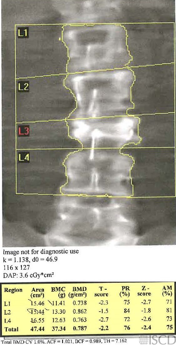
Burst Fracture of L3

There is a compression fracture at L3 on this Hologic lumbar spine DXA scan. L3 was removed from analysis.
This is the accompanying radiograph showing the compression fracture at L3.
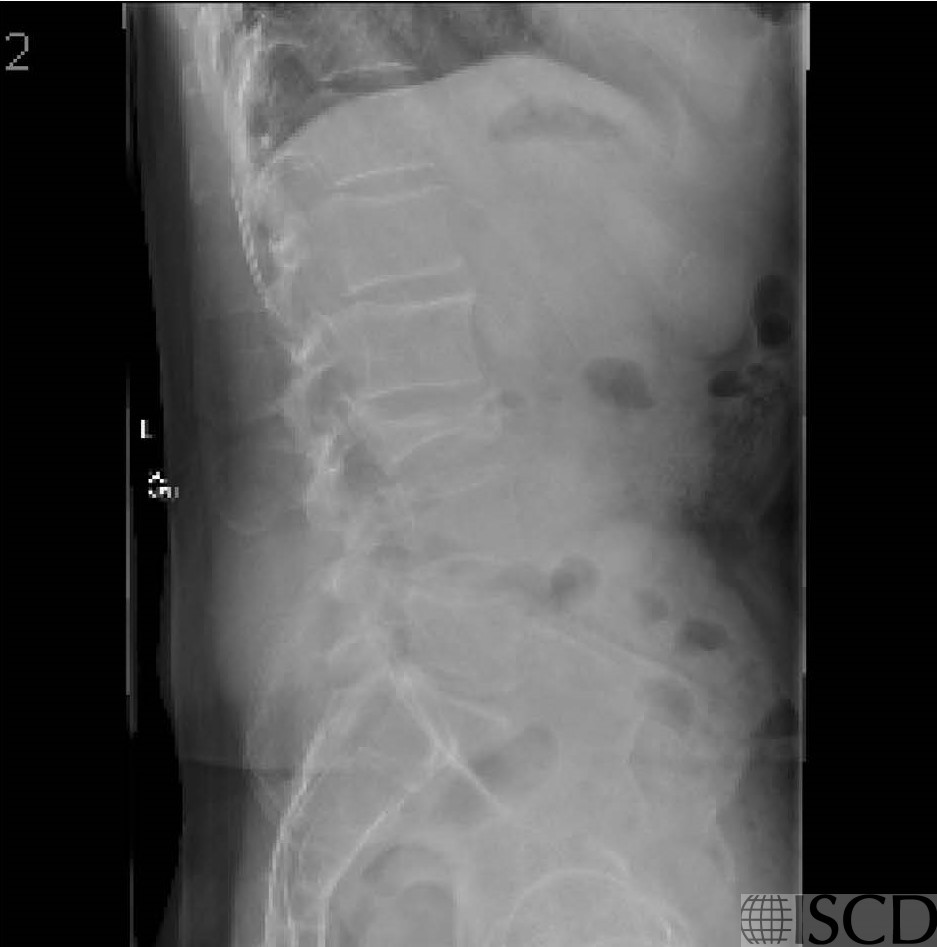
The Hologic DXA scan shows a compression fracture at L3. The accompanying lateral radiograph shows a L3 compression fracture. The fracture was a burst compression fracture.
Sarah L Morgan, MD, RD, CCD, The University of Alabama at Birmingham
• Martineau, P., S. Bazarjani, and L.S. Zuckier, Artifacts and Incidental Findings Encountered on Dual-Energy X-Ray Absorptiometry: Atlas and Analysis. Semin Nucl Med, 2015. 45(5): p. 458-69.
• Bazzocchi, A., et al., Incidental findings with dual-energy X-ray absorptiometry: spectrum of possible diagnoses. Calcif Tissue Int, 2012. 91(2): p. 149-56
• Lentle, B., J. Trollip, and K. Lian, The Radiology of Osteoporotic Vertebral Fractures Redux. J Clin Densitom, 2016. 19(1): p. 40-7.
• Oei, L., et al., Osteoporotic Vertebral Fractures as Part of Systemic Disease. J Clin Densitom, 2016. 19(1): p. 70-80.
• Ryan, P.J., et al., The effect of vertebral collapse on spinal bone mineral density measurements in osteoporosis. Bone Miner, 1992. 18(3): p. 267-72.
• McKiernan, F.E., Atypical femoral diaphyseal fractures documented by serial DXA. J Clin Densitom, 2010. 13(1): p. 102-3.
• McKiernan, F.E., et al., A long femur scan field does not alter proximal femur bone mineral density measurements by dual-energy X-ray absorptiometry. J Clin Densitom, 2011. 14(3): p. 354-8.
• Prater, G.L., et al., The effect of extending femur scan length on BMD results on the Hologic Discovery-W scanner. J Clin Densitom, 2014. 17(4): p. 518-21.
• Morgan, S.L., et al., Nonprogression of vertebral area or bone mineral content on DXA does not predict compression fractures. J Clin Densitom, 2006. 9(3): p. 261-4.
• Adler, R.A., R.R. Nordlie, and T.S. Burke, Increased bone mineral density in a man with known compression fractures. J Clin Densitom, 1998. 1(3): p. 275-8.