Calcinosis

The image shows a left proximal femur scan from a patient with calcinosis from scleroderma.

The image shows a right proximal femur scan from a patient with calcinosis from scleroderma.
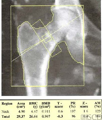
The DXA scan shows calcinosis in a patient with known scleroderma. Calcinosis is the presence of calcium deposits in soft tissues. The differential diagnosis of calcinosis includes infections, tumors, connective tissue disease, hyperphosphatemia and hypercalcemia. The calcinosis over bone will overestimate bone mineral density. The calcinosis in the soft tissues will be mapped and deleted from the soft tissue baseline, and this could be confirmed by evaluation of the undo view on a Hologic scan.
Sarah L Morgan, MD, RD, CCD, The University of Alabama at Birmingham
• Downing, M.R., et al., Impact of trochanteric heterotopic ossification on measurement of femoral bone density following cemented total hip replacement. J Orthop Res, 2008. 26(10): p. 1334-9.
• Jaovisidha, S., et al., Influence of heterotopic ossification of the hip on bone densitometry: a study in spinal cord injured patients. Spinal Cord, 1998. 36(9): p. 647-53.
• Okano, K., et al., Bone mineral density is not related to heterotopic ossification after total hip arthroplasty. Int Orthop, 2012. 36(6): p. 1163-6.
• Schneider, D.L., Pitfalls in interpretation: calcium that’s not bone. J Clin Densitom, 1998. 1(4): p. 405-6.
• Barfield, W.R., R.E. Holmes, and L.A. Hartsock, Heterotopic Ossification in Trauma. Orthop Clin North Am, 2017. 48(1): p. 35-46.
• Dey, D., et al., The traumatic bone: trauma-induced heterotopic ossification. Transl Res, 2017. 186: p. 95-111.
• Ohlmeier, M., et al., Muscle localization of heterotopic ossification following spinal cord injury. Spine J, 2017.
• Stewart, C.A., et al., Heterotopic ossification. Effect on dual-photon absorptiometry of the hip. Clin Nucl Med, 1990. 15(10): p. 697-700.