Celiac Disease

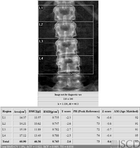
This is the lumbar spine Hologic DXA scan from a woman with biospy-proven celiac disease.
This is the left proximal femur Hologic DXA scan from a woman with biopsy-proven celiac disease.

This is a DXA scan from a 65-year-old Caucasian female referred with low bone mineral density and a poorly healing fracture. The second cause work up demonstrated celiac sprue with anti-TTG > 150. The diagnosis was subsequently confirmed on a small bowel biopsy. There are no specific DXA finding that would suggest celiac sprue over another secondary cause. A thoughtful secondary cause work up is important in patients being evaluated for low bone mineral density and fracture. Relatively large increases in bone mineral density have been reported with the treatment of celiac sprue with a gluten-free diet.
Sarah L Morgan, MD, RD, CCD, The University of Alabama at Birmingham
• Duerksen, D.R., M. Ali, and W.D. Leslie, Dramatic effect of vitamin D supplementation and a gluten-free diet on bone mineral density in a patient with celiac disease. J Clin Densitom, 2012. 15(1): p. 120-3.
• Duerksen, D.R. and W.D. Leslie, Longitudinal evaluation of bone mineral density and body composition in patients with positive celiac serology. J Clin Densitom, 2011. 14(4): p. 478-83.
• Kavak, U.S., et al., Bone mineral density in children with untreated and treated celiac disease. J Pediatr Gastroenterol Nutr, 2003. 37(4): p. 434-6.
• Xing, Y. and S.L. Morgan, Celiac disease and metabolic bone disease. J Clin Densitom, 2013. 16(4): p. 439-44.
• Rabelink, N.M., et al., Bone pain and extremely low bone mineral density due to severe vitamin D deficiency in celiac disease. Arch Osteoporos, 2011. 6: p. 209-13.
• Micic, D., V.L. Rao, and C.E. Semrad, Celiac Disease and Its Role in the Development of Metabolic Bone Disease. J Clin Densitom, 2020. 23(2): p. 190-199.