Femoral Neck Region of Interest Positioning/ Hologic
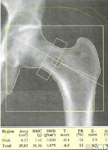
In this left Hologic hip scan, the femoral neck box is incorrectly placed. It should be anchored to the greater trochanter. The width of the femoral neck box should be 15 pixels.

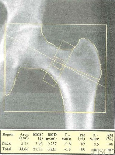
The same Hologic hip scan was altered to correctly position the femoral neck box – anchored to the greater trochanter.

In the left panel, the femoral neck box is incorrectly placed for a Hologic hip scan. On a Hologic scanner, the femoral neck box should be anchored to the greater trochanter. The right panel shows correct positioning of the femoral neck box on a Hologic scan, where the femoral neck box is anchored to the greater trochanter. There should be 3 corners of the femoral neck box in soft tissue.
Sarah L Morgan, MD, RD, CCD, The University of Alabama at Birmingham