Follow-up Hip Scan with the Lesser Trochanter Incompletely Outlined
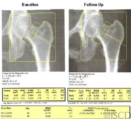
The left panel shows the baseline scan and the right panel shows the follow up scan with the lesser trochanter not outlined.

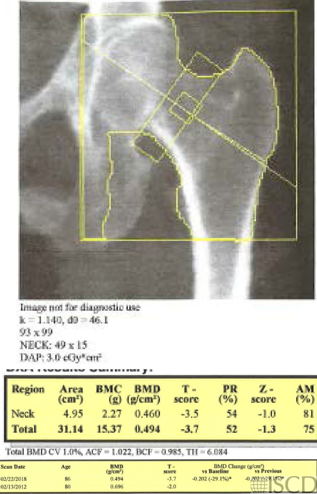
This panel shows the corrected scan with the ischium deleted in approximately the same way and the lesser trochanter is now outlined.

The baseline scan is presented in the upper left panel. In the upper right panel is the follow-up scan. The ischium was omitted differently, and the lesser trochanter was not outlined. The bottom panel shows the corrected scan with the ischium omitted in approximately the same way and the lesser trochanter now outlined. The total hip bone mineral density is higher with the lesser trochanter outlined. The scans were 6 years apart and there is a significant difference at the total hip with both scenarios, however the magnitude of the loss is much greater without the lesser trochanter being outlined.
Sarah L Morgan, MD, RD, CCD, The University of Alabama at Birmingham