Hologic Total Hip Region of Interest Box Incorrect

In this panel, the Hologic analysis of the hip is incorrect. The bottom of the ROI box is down > 10 pixels from the lesser trochanter. The default width of the femoral neck is incorrect, and the default width is 15 pixels. The correct default width is 15 pixels. On the Hologic printout, the size of the hip global ROI and femoral neck are present below the image.

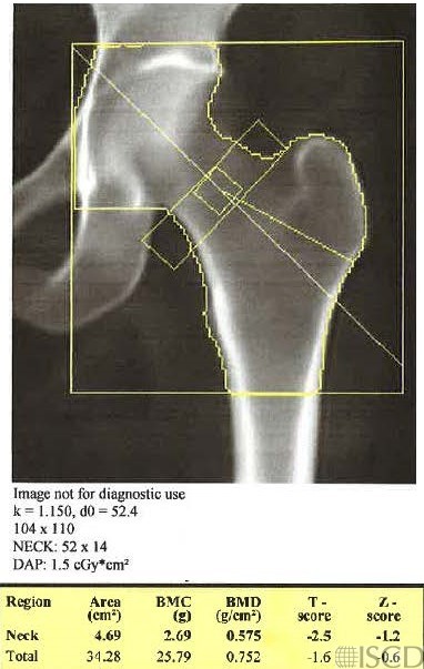
The outside facility submitted a corrected image, but the femoral neck box is now 14 pixels wide instead of 15 pixels wide. The bottom of the global hip ROI box is now 10 pixels below the lesser trochater. Notice that the total hip bone mineral density is now lower because less cortical bone is included.
The left panel shows an incorrect analysis of the left proximal femur from an outside facility. The bottom of the hip region of interest box is down > 10 pixels from the bottom of the lesser trochanter. In addition, the default size of the femoral neck is incorrect. After a call to the facility, explaining the issues, the panel on the right shows a corrected scan from the outside facility. However, the femoral neck default width is still incorrect. A tiny portion of the femoral neck box is overlying the ischium, however, the ischium has been painted out. Notice that by bringing the bottom of the box up, the total hip bone mineral density declines, since less cortical bone is included.
Sarah L Morgan, MD, RD, CCD, The University of Alabama at Birmingham
• Feit, A., et al., Effect of positioing of the region of interest on bone density of the hip. . J Clin Densitom, 2020: 23(3) p 426-431.
• Morgan, S.L. and F. Peace, Do changes in the femoral neck box size make significant difference in femoral neck BMD? . J Clin Densitom, 2011. 14 p. 156
• McKiernan, F.E., et al., A long femur scan field does not alter proximal femur bone mineral density measurements by dual-energy X-ray absorptiometry. J Clin Densitom, 2011. 14(3): p. 354-8.
• Prater, G.L., et al., The effect of extending femur scan length on BMD results on the Hologic Discovery-W scanner. J Clin Densitom, 2014. 17(4): p. 518-21.
• Celik, O., et al., The effect of hip rotation on bone mineral density of the proximal femur measured by dual energy X-ray absorptiometry. Eklem Hastalik Cerrahisi, 2009. 20(2): p. 71-7.
• Cheng, X.G., et al., Effects of anteversion on femoral bone mineral density and geometry measured by dual energy X-ray absorptiometry: a cadaver study. Bone, 1997. 21(1): p. 113-7.
• Girard, M.S., et al., Measured femoral density by dual-energy X-ray absorptiometry as a function of rotation. Orthop Rev, 1994. 23(1): p. 38-40.
• Goh, J.C., S.L. Low, and K. Bose, Effect of femoral rotation on bone mineral density measurements with dual energy X-ray absorptiometry. Calcif Tissue Int, 1995. 57(5): p. 340-3.
• Lekamwasam, S. and R.S. Lenora, Effect of leg rotation on hip bone mineral density measurements. J Clin Densitom, 2003. 6(4): p. 331-6.
• Rosenthall, L., Range of change of measured BMD in the femoral neck and total hip with rotation in women. J Bone Miner Metab, 2004. 22(5): p. 496-9.
• Tang, H., S.M. Ren, and X.Z. Luo, [Effect of femoral rotation on hip bone mineral density measurement]. Zhongguo Yi Xue Ke Xue Yuan Xue Bao, 2003. 25(3): p. 267-70.
• Wilson, C.R., et al., The effect of positioning on dual energy X-ray bone densitometry of the proximal femur. Bone Miner, 1991. 13(1): p. 69-76.
• McKiernan, F. and W. Washington, Effect of subtle positioning flaws on measured bone mineral density of the hip. J Clin Densitom, 2005. 8(3): p. 330-4.
• Hans, D., et al., Effects of a new positioner on the precision of hip bone mineral density measurements. J Bone Miner Res, 1997. 12(8): p. 1289-94.