Inaccurately Elevated T-scores on DXA Scans in a Patient with Metastatic Prostate Cancer
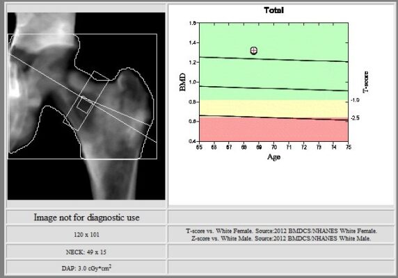
This image shows DXA of the left hip. The left total hip bone mineral density is 1.316 grams per square centimeter, with elevated T-score of 3.1. There are diffuse heterogeneous sclerotic changes.

This table summarizes the results of DXA of the left hip.

This image shows DXA of the lumbar spine. The L1-L4 bone mineral density is 1.728 grams per square centimeter, with elevated T-score of 6.2. There are diffuse heterogeneous sclerotic changes.

This table summarizes the results of DXA of the lumbar spine.

The Hologic lumbar spine and left total hip DXA scans demonstrated elevated T scores in a patient with metastatic prostate cancer due to diffuse heterogeneous sclerotic changes. In the setting of skeletal metastatic disease, bone mineral density (BMD) measurements can be artificially increased, leading to inaccurately elevated T-scores. When unusually high BMD values are observed on DXA, results should be interpreted with caution and potential contributions from artifacts, generalized skeletal pathology and focal lesions should be considered.
Tam Dinh, MD, University of New Mexico, Internal Medicine Department.
Vijayalakshmi Kumar, MD, New Mexico VA Health Care System, Rheumatology Department.
1. Paccou J, Michou L, Kolta S, Debiais F, Cortet B, Guggenbuhl P. High bone mass in adults. Joint Bone Spine. 2018 Dec;85(6):693-699. doi: 10.1016/j.jbspin.2018.01.007. Epub 2018 Mar 2. PMID: 29407041.
2. D’Oronzo S, Cives M, Lauricella E, Stucci S, Centonza A, Gentile M, Ostuni C, Porta C. Assessment of bone turnover markers and DXA parameters to predict bone metastasis progression during zoledronate treatment: a single-center experience. Clin Exp Med. 2024 Jan 19;24(1):7. doi: 10.1007/s10238-023-01280-1. PMID: 38240866; PMCID: PMC10798926.
3. Forbes V, Taxel P. Onset of asymptomatic skeletal metastatic disease seen on DXA. AACE Clin Case Rep. 2018;4(6):e472–5