Incorrect Femoral Neck Positioning/Hologic

In this Hologic left hip analysis the placement of the femoral neck box is incorrect. The femoral neck box contains a small portion of the greater trochanter.
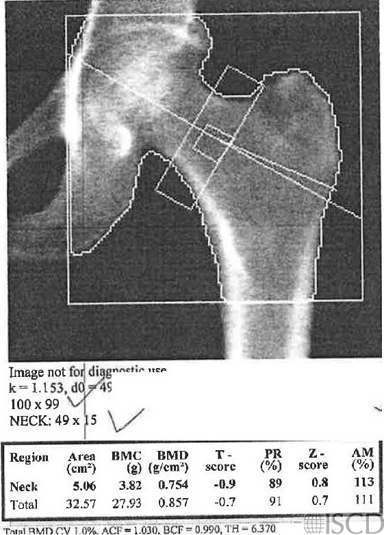
This is the corrected analysis with the femoral neck box now anchored to the greater trochanter.

In a Hologic hip analysis, the femoral neck should be anchored to the greater trochanter. In the image on the left, there is a small amount of greater trochanter within the femoral neck box. The corrected analysis is seen on the right, where the femoral neck box is anchored to the left greater trochanter. While the numerical difference is not great, it is important to perform a correct analysis every time. This is important for both accuracy and precision.
Sarah L Morgan, MD, RD, CCD, The University of Alabama at Birmingham
• Feit, A., et al., Effect of positioning of the region of interest on bone density of the hip. . J Clin Densitom, 2020: 23(3) p 426-431.