Mastocytosis

There is diffuse heterogeneous sclerosis in the hip scan and the bone mineral density is elevated
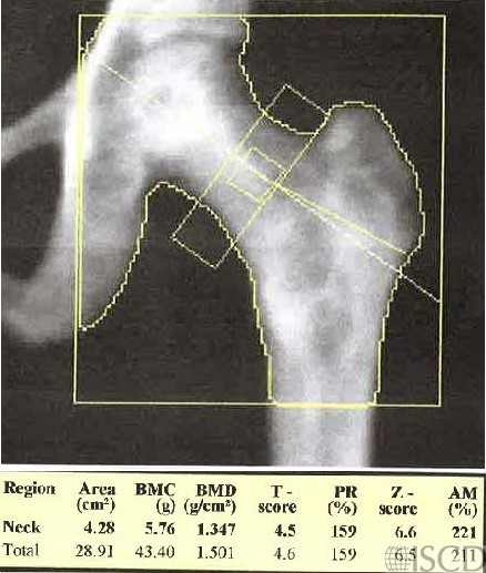
This is the left proximal Hologic femur scan of a patient with known mastocytosis. The chest, abdomen, and pelvis CT scans demonstrated diffuse heterogeneous sclerosis of the bones.
Sarah L Morgan, MD, RD, CCD, The University of Alabama at Birmingham
• Johansson, C., et al., Bone density, bone markers and bone radiological features in mastocytosis. Age Ageing, 1996. 25(1): p. 1-7.
• Rossini, M., et al., Bone mineral density, bone turnover markers and fractures in patients with indolent systemic mastocytosis. Bone, 2011. 49(4): p. 880-5.
• Artuso, A., et al., Longitudinal Evaluation of Bone Mineral Density and Bone Metabolism Markers in Patients with Indolent Systemic Mastocytosis Without Osteoporosis. Calcif Tissue Int, 2017. 100(1): p. 40-46.
• Zechner, C. and U. Gruntmanis, Systemic Mastocytosis With Decreased Bone Density and Fractures. Mayo Clin Proc, 2015. 90(6): p. 843-4.
• Whyte, M.P., Misinterpretation of osteodensitometry with high bone density: BMD Z > or = + 2.5 is not “normal”. J Clin Densitom, 2005. 8(1): p. 1-6.