Spine Positioning and Rotation

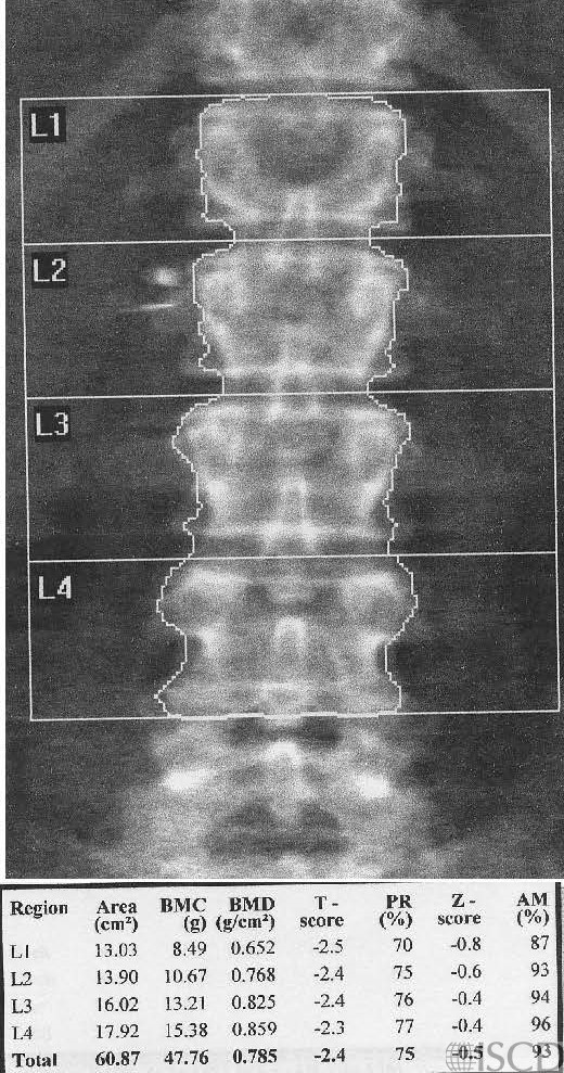
Baseline Hologic lumbar spine scan showing clips lateral to L2 on the right.
Follow-up Hologic lumbar spine scan with the spine rotated with clips projecting over L2. The spine is also not straight.
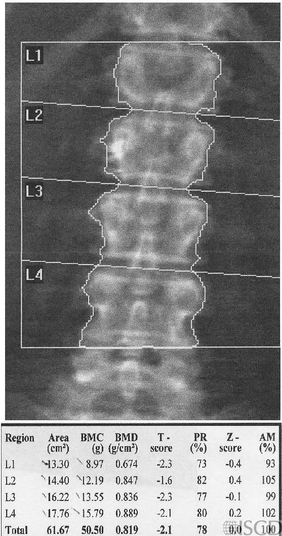
The artifacts lateral to L2 on the left in the baseline scan can be used to gauge a difference in spine positioning on the followup scan. On the follow-up scan, the clips now project over L2, meaning that L2 should be removed from analysis. For optimal precision, the positioning of the patient should be analogous between baseline and follow-up scans. Differences in axial rotation of the spine have been studied by several authors, with a variety of spine phantoms. Generally, lateral and axial rotation of the spine increases the area, with a corresponding decrease in bone density.
Sarah L Morgan, MD, RD, CCD, The University of Alabama at Birmingham
• Cetin, A., et al., Evaluation of the patient positioning during DXA measurements in daily clinical practice. Clin Rheumatol, 2008. 27(6): p. 713-5.
• Cheng, J.C., et al., The effect of vertebral rotation of the lumbar spine on dual energy X-ray absorptiometry measurements: observational study. Hong Kong Med J, 2001. 7(3): p. 241-5.
• Izadyar, S., et al., The Effect of the Lumbar Vertebral Malpositioning on Bone Mineral Density Measurements of the Lumbar Spine by Dual-Energy X-Ray Absorptiometry. J Clin Densitom, 2016. 19(3): p. 277-81.
• Jeon, Y.K., et al., Effect of increased axial rotation angle on bone mineral density measurements of the lumbar spine. Spine J, 2014. 14(9): p. 2150-4.
• Watts, N.B., Fundamentals and pitfalls of bone densitometry using dual-energy X-ray absorptiometry (DXA). Osteoporos Int, 2004. 15(11): p. 847-54.
• Choplin R.H., Lenchik L and S. Wuertzer, A practical approach to interpretation of Dual-Energy X-ray Absorptiometry (DXA) for assessment of bone density. . Curr Radiol Rep 2014. 2(48).
• Dasher, L.G., C.D. Newton, and L. Lenchik, Dual X-ray absorptiometry in today’s clinical practice. Radiol Clin North Am, 2010. 48(3): p. 541-60.
• Theodorou, D.J. and S.J. Theodorou, Dual-energy X-ray absorptiometry in clinical practice: application and interpretation of scans beyond the numbers. Clin Imaging, 2002. 26(1): p. 43-9.
• Mergler, S., et al., Lumbar spine and total-body dual-energy X-ray absorptiometry in children with severe neurological impairment and intellectual disability: a pilot study of artefacts and disrupting factors. Pediatr Radiol, 2012. 42(5): p. 574-83.
• Choi, J.S., The influence of soft tissue recognition erros on BMD value-A case report: Recipient of Young Investigator Award J Clin Densitom, 2012. 15(4): p. 483
• Fuleihan, G.E., et al., Reproducibility of DXA absorptiometry: a model for bone loss estimates. J Bone Miner Res, 1995. 10(7): p. 1004-14.
• Fuerst, T., et al., Quality Assurance in Bone Densitometry in Bone Densitometry and Osteoporosis H.K. Genant, G. Guglielmi, and M. Jergas, Editors. 1998, Springer Berlin.
• Hansen, K., et al., DXA Errors are Common and Likely Adversely Affect Clinical Care: DXA Quality Improvement is Needed. J Bone Miner Res 2016. 31((Suppl 1) Available at http://www.asbmr.org/ItineraryBuilder/Presentation Detail.aspx?pid=83c01c31-237b-4f07-81a5-1eeb2a7968aa&ptag=AuthorDetail&aid=00000000-0000-0000-0000-000000000000. ).
• Promma, S., et al., Errors in Patient Positioning for Bone Mineral Density Assessment by Dual x-ray Absorptiometry: Effect of Technologist Retraining. J Clin Densitom, 2018. 21(2): p. 252-259.
• Cetin, A., et al., Evaluation of the patient positioning during DXA measurements in daily clinical practice. Clin Rheumatol, 2008. 27(6): p. 713-5.
• Staron, R.B., et al., Computerized bone densitometric analysis: operator-dependent errors. Radiology, 1999. 211(2): p. 467-70.
• Baniak, N., S. Grzybowski, and W.P. Olszynski, Dual-energy x-ray absorptiometry scan autoanalysis vs manual analysis. J Clin Densitom, 2014. 17(1): p. 97-103.