Total Hip Region of Interest Incorrect/Hologic

The bottom of the total hip region of interest is incorrect on this Hologic scan. The bottom line is placed 10 pixels below the lesser trochanter. This analysis would overestimate the bone mineral density in the total hip because more cortical bone in the femoral shaft is included the the total hip region of interest.
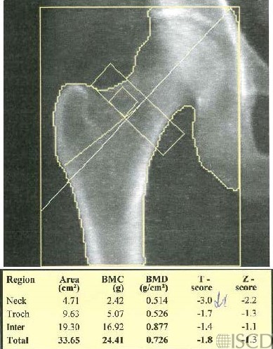
The bottom of the hip region of interest box is down too low. On a Hologic scan, the bottom part of the box should be 10 pixels below the bottom of the lesser trochanter. This positioning will tend to overestimate the bone mineral density in the total hip. The distance between the blue and yellow lines is 5 pixels. The upper, inner and outer edges are all set with a blue line that “touches” bone and the yellow line that is 5 pixels out. The exception is the bottom line, it is 10 lines pixels below.
Sarah L Morgan, MD, RD, CCD, The University of Alabama at Birmingham
• Feit, A., et al., Effect of positioing of the region of interest on bone density of the hip. . J Clin Densitom, 2020: 23(3) p 426-431.
• Morgan, S.L. and F. Peace, Do changes in the femoral neck box size make significant difference in femoral neck BMD? . J Clin Densitom, 2011. 14 p. 156
• McKiernan, F.E., et al., A long femur scan field does not alter proximal femur bone mineral density measurements by dual-energy X-ray absorptiometry. J Clin Densitom, 2011. 14(3): p. 354-8.
• Prater, G.L., et al., The effect of extending femur scan length on BMD results on the Hologic Discovery-W scanner. J Clin Densitom, 2014. 17(4): p. 518-21.
• Celik, O., et al., The effect of hip rotation on bone mineral density of the proximal femur measured by dual energy X-ray absorptiometry. Eklem Hastalik Cerrahisi, 2009. 20(2): p. 71-7.
• Cheng, X.G., et al., Effects of anteversion on femoral bone mineral density and geometry measured by dual energy X-ray absorptiometry: a cadaver study. Bone, 1997. 21(1): p. 113-7.
• Girard, M.S., et al., Measured femoral density by dual-energy X-ray absorptiometry as a function of rotation. Orthop Rev, 1994. 23(1): p. 38-40.
• Goh, J.C., S.L. Low, and K. Bose, Effect of femoral rotation on bone mineral density measurements with dual energy X-ray absorptiometry. Calcif Tissue Int, 1995. 57(5): p. 340-3.
• Lekamwasam, S. and R.S. Lenora, Effect of leg rotation on hip bone mineral density measurements. J Clin Densitom, 2003. 6(4): p. 331-6.
• Rosenthall, L., Range of change of measured BMD in the femoral neck and total hip with rotation in women. J Bone Miner Metab, 2004. 22(5): p. 496-9.
• Tang, H., S.M. Ren, and X.Z. Luo, [Effect of femoral rotation on hip bone mineral density measurement]. Zhongguo Yi Xue Ke Xue Yuan Xue Bao, 2003. 25(3): p. 267-70.
• Wilson, C.R., et al., The effect of positioning on dual energy X-ray bone densitometry of the proximal femur. Bone Miner, 1991. 13(1): p. 69-76.
• McKiernan, F. and W. Washington, Effect of subtle positioning flaws on measured bone mineral density of the hip. J Clin Densitom, 2005. 8(3): p. 330-4.
• Hans, D., et al., Effects of a new positioner on the precision of hip bone mineral density measurements. J Bone Miner Res, 1997. 12(8): p. 1289-94.