Aortic calcification on spine DXA

Figure 1 – DXA of left hip and lumbar spine.
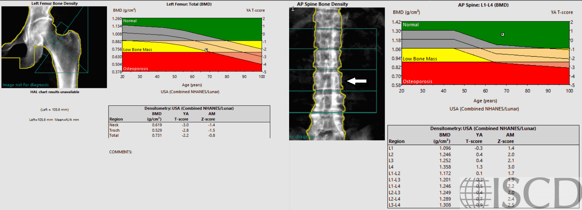
Figure 2 – X-rays of lumbar spine showing dense aortoiliac calcification.
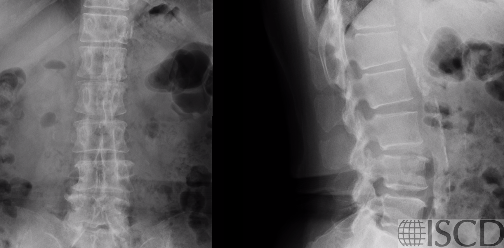
Figure 3 – CT showing dense aortoiliac calcification.

Case Description:
68 year old woman: Baseline BMD assessment, no osteoporosis risk factors. Treated hypertension and hyperlipidemia.
The patient has osteoporosis (minimum T-score -3.0 femur neck). The spine BMD is artifactually elevated by dense aortic calcification that superimposes on the left side of the lumbar spine, see arrow (T-score +0.5). All vertebral levels are affected — there is no saving this spine!
Credit: William D. Leslie, MD FRCPC MSc CCD, University of Manitoba