Clips

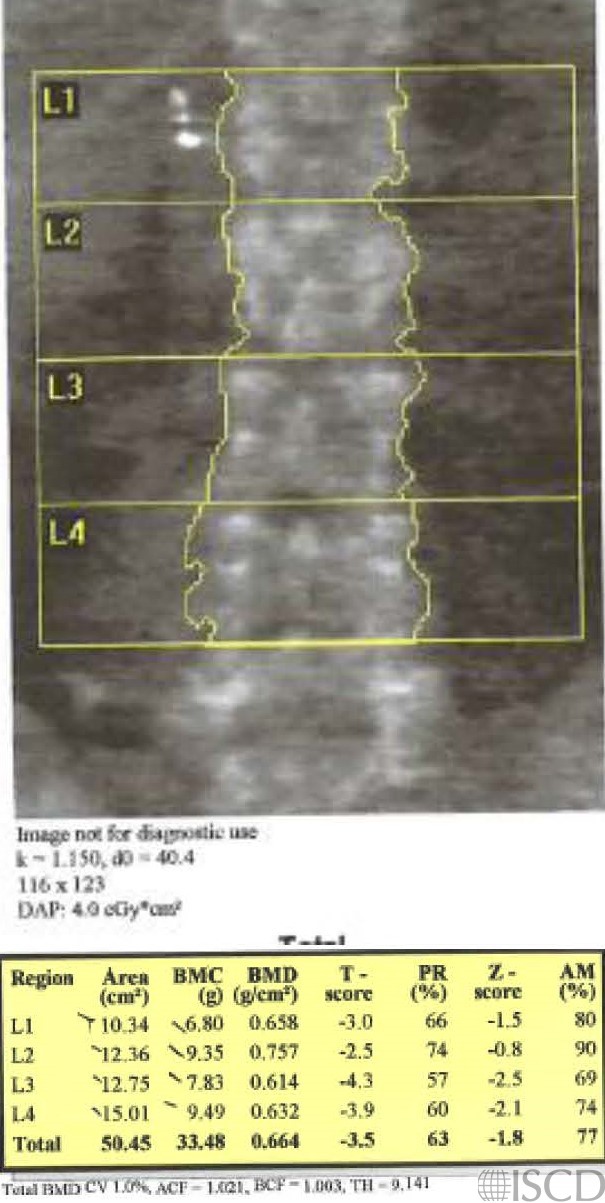
This Hologic spine images shows artifacts lateral to L1 on the right that are cholecystectomy clips.
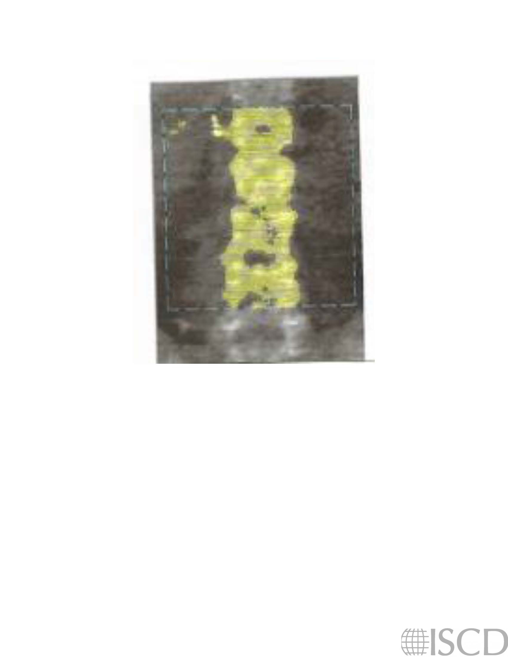
The undo of the image shows that the clips are recognized and would be omitted by the software from the soft tissue baseine
There are metallic artifacts lateral to L1 on the right that were from a cholecystectomy. Gall bladder clips are white on a dual-energcy Hologic scan, as opposed to tantalum clips, which are black on the dual-energy image.
Sarah L Morgan, MD, RD, CCD, The University of Alabama at Birmingham
• Martineau, P., S. Bazarjani, and L.S. Zuckier, Artifacts and Incidental Findings Encountered on Dual-Energy X-Ray Absorptiometry: Atlas and Analysis. Semin Nucl Med, 2015. 45(5): p. 458-69.
• Bazzocchi, A., et al., Incidental findings with dual-energy X-ray absorptiometry: spectrum of possible diagnoses. Calcif Tissue Int, 2012. 91(2): p. 149-56.
• Bojinca, V., D. Opris, and M. Bonjinca, Artifacts and pitfalls in DXA scan images and interpretation. J Clin Densitom, 2012. 15(4): p. 486-487.