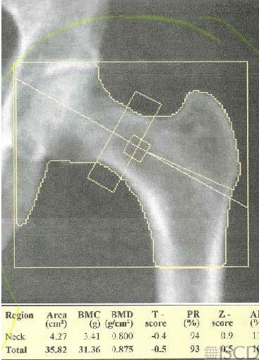
Femoral Neck Region of Interest Positioning/ Hologic


In this left Hologic hip scan, the femoral neck box is incorrectly placed. It should be anchored to the greater trochanter. The width of the femoral neck box should be 15 pixels.

The same Hologic hip scan was altered to correctly position the femoral neck box – anchored to the greater trochanter.

Case Description:

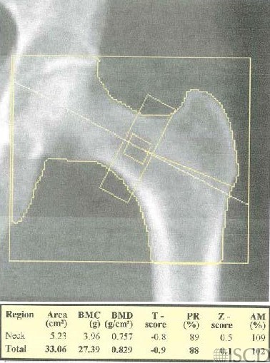
In the left panel, the femoral neck box is incorrectly placed for a Hologic hip scan. On a Hologic scanner, the femoral neck box should be anchored to the greater trochanter. The right panel shows correct positioning of the femoral neck box on a Hologic scan, where the femoral neck box is anchored to the greater trochanter.  There should be 3 corners of the femoral neck box in soft tissue.

Credit:

Sarah L Morgan, MD, RD, CCD, The University of Alabama at Birmingham

References:
  • McKiernan, F.E., et al., A long femur scan field does not alter proximal femur bone mineral density measurements by dual-energy X-ray absorptiometry. J Clin Densitom, 2011. 14(3): p. 354-8.
  • Prater, G.L., et al., The effect of extending femur scan length on BMD results on the Hologic Discovery-W scanner. J Clin Densitom, 2014. 17(4): p. 518-21.
  • Celik, O., et al., The effect of hip rotation on bone mineral density of the proximal femur measured by dual energy X-ray absorptiometry. Eklem Hastalik Cerrahisi, 2009. 20(2): p. 71-7.
  • Cheng, X.G., et al., Effects of anteversion on femoral bone mineral density and geometry measured by dual energy X-ray absorptiometry: a cadaver study. Bone, 1997. 21(1): p. 113-7.
  • Girard, M.S., et al., Measured femoral density by dual-energy X-ray absorptiometry as a function of rotation. Orthop Rev, 1994. 23(1): p. 38-40.
  • Goh, J.C., S.L. Low, and K. Bose, Effect of femoral rotation on bone mineral density measurements with dual energy X-ray absorptiometry. Calcif Tissue Int, 1995. 57(5): p. 340-3.
  • Lekamwasam, S. and R.S. Lenora, Effect of leg rotation on hip bone mineral density measurements. J Clin Densitom, 2003. 6(4): p. 331-6.
  • Rosenthall, L., Range of change of measured BMD in the femoral neck and total hip with rotation in women. J Bone Miner Metab, 2004. 22(5): p. 496-9.
  • Tang, H., S.M. Ren, and X.Z. Luo, [Effect of femoral rotation on hip bone mineral density measurement]. Zhongguo Yi Xue Ke Xue Yuan Xue Bao, 2003. 25(3): p. 267-70.
  • Wilson, C.R., et al., The effect of positioning on dual energy X-ray bone densitometry of the proximal femur. Bone Miner, 1991. 13(1): p. 69-76.
  • McKiernan, F. and W. Washington, Effect of subtle positioning flaws on measured bone mineral density of the hip. J Clin Densitom, 2005. 8(3): p. 330-4.
  • Hans, D., et al., Effects of a new positioner on the precision of hip bone mineral density measurements. J Bone Miner Res, 1997. 12(8): p. 1289-94.
  • Feit, A., et al., Effect of positioing of the region of interest on bone density of the hip. . J Clin Densitom, 2020: 23(3) p 426-431.